Calcaneus (heel bone)
The calcaneus is the largest tarsal bone forming the heel. It transmits weight, anchors the Achilles tendon and plantar fascia, and is important in gait. Anatomy, function and clinical notes.
The calcaneus is the large bone at the back of the foot that forms the prominence of the heel and helps transfer body weight to the ground. It is one of the tarsal bones and works with neighboring bones and soft tissues to support standing, walking and running. For general context about the foot's structure see related overview. The name derives from Latin; alternative forms include calcaneum or calcaneus and relate to the meaning "heel" (etymology).
Image gallery
10 Images


Anatomical features
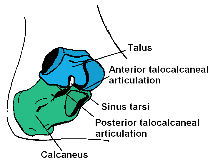
The calcaneus has a robust, irregular shape with several recognizable landmarks. Posteriorly it carries the calcaneal tuberosity, the broad surface that receives the insertion of the Achilles (calcaneal) tendon. Superiorly it presents articular facets for the talus that form part of the subtalar joint. Anteriorly it articulates with the cuboid bone. Medially, a shelf-like projection called the sustentaculum tali supports the talus and provides a pulley surface for the flexor hallucis longus tendon. The plantar surface gives attachment to the plantar aponeurosis and several intrinsic foot muscles.
Function and biomechanics
Functionally the calcaneus transmits load from the tibia and talus to the ground and acts as a lever for the triceps surae (calf) muscles via the Achilles tendon, amplifying plantarflexion force. The subtalar joint between calcaneus and talus allows inversion and eversion of the foot, important for adapting to uneven surfaces. Its shape and soft tissue attachments are central to arch mechanics and shock absorption during gait.
Clinical significance
- Calcaneal fractures occur from high-energy axial loads and can impair gait and ankle alignment; they often require careful orthopedic assessment.
- Insertional Achilles tendinopathy and plantar fasciitis involve structures attached to the calcaneus and are common causes of heel pain.
- Deformities of the calcaneus—congenital or acquired—affect foot posture and may alter walking mechanics.
Development and comparative notes
The calcaneus develops as part of the tarsal skeleton during early life and remodels in response to mechanical load. It is the largest tarsal bone in humans, reflecting the bipedal requirement to bear body weight on the hindfoot. In comparative anatomy the heel region shows variation across vertebrates correlated with differences in locomotion.
Understanding the calcaneus—its form, attachments and articulations—helps explain many common foot conditions and guides surgical or rehabilitative approaches to heel injuries and pain.
Related articles
Author
AlegsaOnline.com Calcaneus (heel bone) Leandro Alegsa
URL: https://en.alegsaonline.com/art/16029