Santiago Ramón y Cajal
Spanish physician and histologist (1852–1934) celebrated for founding modern neuroscience, the neuron doctrine, and for detailed silver-stain illustrations of brain cells and circuits.
Overview
Santiago Ramón y Cajal (1 May 1852 – 18 October 1934) was a Spanish physician, histologist and anatomist whose microscopic studies transformed ideas about the nervous system. He shared the 1906 Nobel Prize in Physiology or Medicine with Camillo Golgi for complementary work on neural tissue and staining techniques that revealed cellular detail in the brain (nervous system).
Image gallery
10 Images


Techniques and discoveries
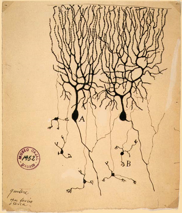
Using the silver (Golgi) staining method, Cajal examined thin sections of nervous tissue mounted on microscope slides. Rather than inventing the stain, he adapted and refined its use to reveal individual cells and their processes. From these observations he argued that the nervous system is made of discrete cells, a concept often called the neuron doctrine. This idea challenged the prevailing reticular theory, which held that neural tissue formed a continuous network.

Illustration and description of structures
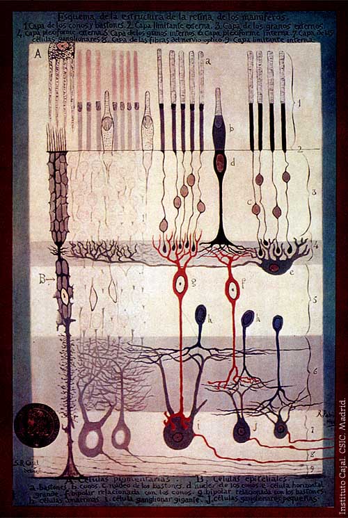
Cajal combined careful microscopy with exceptional draftsmanship, producing hundreds of drawings that clarify cell types and circuits. His illustrations include detailed depictions of hippocampal circuitry, layers of the cerebellum, optic centers and the retina. Examples commonly reproduced in texts show the rodent hippocampus, avian cerebellar cells and sections through the optic tectum.

Major works and influence
Across monographs and many papers he described neuronal morphology, the direction of nerve impulses along axons and the specialization of dendritic arbors. His major compilations of neural structure influenced generations of anatomists and physiologists, and his images remain pedagogical standards for teaching brain organization and development. The study of the mammalian retina and other sensory regions benefited greatly from his systematic approach.

Uses, examples and lasting importance
Cajal's work provided the morphological foundation for modern neurobiology, shaping how researchers interpret connectivity, plasticity and pathology. Clinicians and neuroscientists still refer to his descriptions when identifying cell types—such as Purkinje and granule cells in the cerebellum—and when mapping circuits involved in memory, vision and motor control.

Key contributions and notable facts
- Shared the 1906 Nobel Prize with Golgi for complementary discoveries about neural tissue (Camillo Golgi).
- Formulated and defended the neuron doctrine, opposing the reticular view of the nervous system (neuroscience).
- Produced influential drawings of the hippocampus (hippocampus), the chick cerebellum (cerebellum), the optic tectum (optic tectum) and the mammalian retina (retina).

Cajal's combination of careful experimental technique, clear conceptual insight and precise illustration created a lasting legacy: his findings remain a cornerstone of anatomical neuroscience and an entry point for students and researchers learning how brain cells are organized and interconnected.
Author
AlegsaOnline.com Santiago Ramón y Cajal Leandro Alegsa
URL: https://en.alegsaonline.com/art/130485