
Villi (small intestine): structure, function, and clinical relevance
Villi are finger-like projections in the small intestine that expand absorptive surface area. This article covers their anatomy, role in nutrient uptake, regional differences, development, and clinical importance.
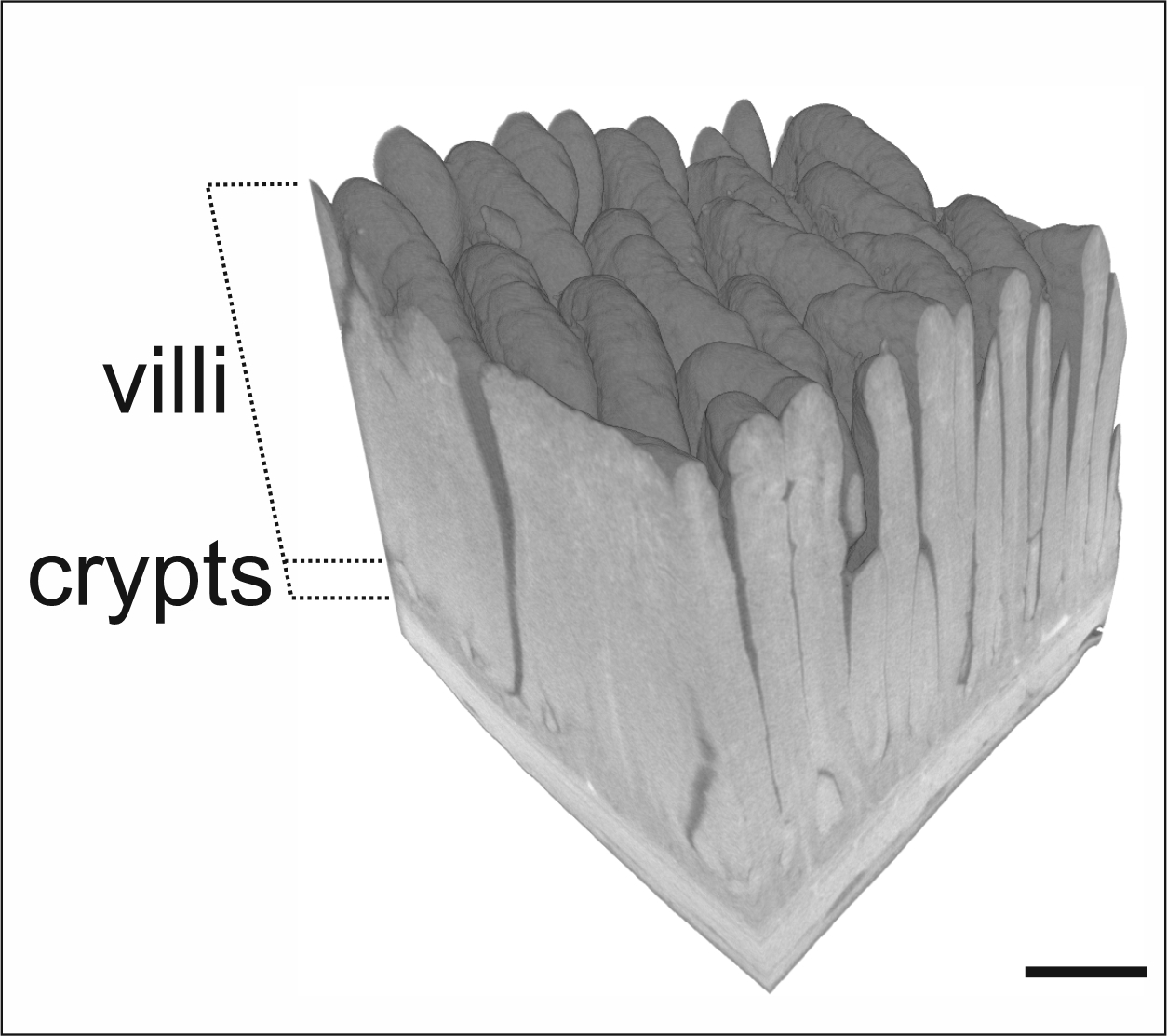
Villi (singular: villus) are microscopic, finger-shaped projections that line the inner surface of the small intestine. They are a central adaptation that increases the absorptive capacity of the gut by enlarging the effective epithelial surface. Each villus is covered by epithelial cells whose apical membranes bear even smaller projections, the microvilli, together forming the intestinal "brush border" that maximizes contact between digested nutrients and the absorbing surface (surface area).
Image gallery
8 Images





Structure and components
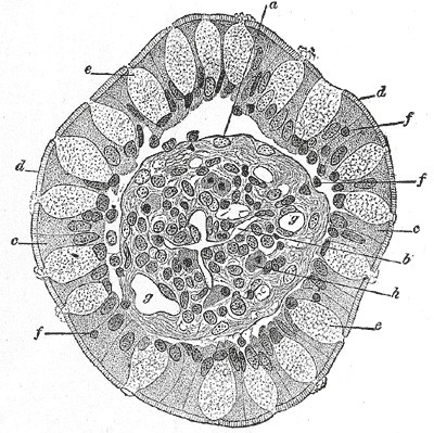
At the gross histological level a villus contains a single layer of enterocytes and interspersed goblet cells resting on a thin connective tissue core (the lamina propria). The lamina propria houses a network of blood capillaries and a central lymphatic vessel called a lacteal. The enterocytes perform absorption and present digestive enzymes at their apical surface. These brush-border enzymes complete the final steps of digestion for certain carbohydrates and peptides.
How villi enable absorption
Before substances reach the villi, larger molecules have been broken down by luminal enzymes such as proteases, lipases and amylases. Within the villus, small water-soluble nutrients — monosaccharides and amino acids — are taken up by enterocytes and pass into nearby capillaries, from where they are carried via the hepatic portal circulation to the liver. Lipid digestion products are reassembled into triglycerides and packaged into chylomicrons that enter the lacteal and then the lymphatic system, eventually reaching the bloodstream. Certain lipid-associated molecules such as cholesterol follow this lymphatic route as well. This division of routes (blood vs lymph) is an important functional distinction.
Regional differences and related structures
The density and morphology of villi vary along the small intestine. Villi are typically longer and more numerous in the jejunum, where most nutrient absorption occurs, and shorter in the ileum. The ileum also contains organized immune structures known as Peyer’s patches that interrupt the villous surface at intervals. Between villi are crypts of Lieberkühn — glandular invaginations where stem cells proliferate and where Paneth cells contribute antimicrobial peptides.
Development, maintenance, and clinical significance
Villi develop during embryogenesis and are maintained by rapid epithelial cell turnover throughout life; stem cells in the crypts continuously renew the villous epithelium. Because of this dynamic nature, the villous surface is vulnerable in several disorders. For example, immune-mediated reactions to certain dietary proteins can produce villous atrophy, reducing absorptive area and causing malabsorption. Resection of segments of the small intestine, inflammatory diseases, and certain infections can also compromise villous function and lead to nutrient deficiencies.
Key points and practical implications
- Villi and microvilli together create a highly folded absorptive surface that facilitates efficient uptake of nutrients and water.
- Different nutrient classes follow distinct pathways after absorption: water-soluble molecules into the blood, lipids largely via lymphatics.
- Healthy villous architecture is essential for nutrition; damage to villi has direct clinical consequences.
- Understanding villus structure informs surgical decisions, treatment of malabsorption syndromes, and nutritional management.
For further basic overviews and diagrams, consult an anatomy or physiology resource on intestinal structure and absorption (absorb, digested food). Additional reading on enzyme function and clinical disorders can be found in resources that discuss digestive enzymes and intestinal pathology.
Related articles
Author
AlegsaOnline.com Villi (small intestine): structure, function, and clinical relevance Leandro Alegsa
URL: https://en.alegsaonline.com/art/105338