
Ventricles of the Brain
Hollow cavities within the brain that produce and circulate cerebrospinal fluid; anatomy, function, development, and clinical relevance explained.
Overview
The brain's ventricles are a connected system of hollow chambers filled with cerebrospinal fluid. Often described together as the ventricular system, they form a continuous space that communicates with the central canal of the spinal cord and plays a central role in protecting and maintaining the brain's internal environment.
Image gallery
7 Images


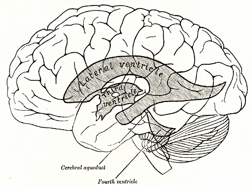

Anatomy and major parts
The ventricular system typically comprises four principal cavities. The two lateral ventricles lie within the cerebral hemispheres and connect to the third ventricle through the interventricular foramina. A narrow channel called the cerebral aqueduct links the third ventricle to the fourth ventricle, which sits between the brainstem and the cerebellum. From there the system continues into the spinal cord via the central canal, described in clinical and anatomical texts as the continuation of the ventricles.
Lining and fluid production
The inner surface of the ventricles is lined by a layer of specialized glial cells called ependyma. Interspersed in ventricular surfaces are tufts of tissue known as the choroid plexuses, where most of the brain's cerebrospinal fluid (CSF) is produced. CSF circulates through the ventricles and over the brain and spinal cord, then is absorbed into the venous system. The properties of this lining influence fluid composition and barrier functions; abnormalities can alter CSF dynamics and pressure.
Functions and clinical importance
Ventricles and their CSF serve several functions: they provide buoyant support that reduces effective brain weight, cushion the brain against mechanical injury, and help remove metabolic waste. Clinically, the ventricular system is central to conditions such as hydrocephalus (excess CSF accumulation), intraventricular hemorrhage, and infections that involve CSF. Imaging modalities like CT and MRI evaluate ventricular size and shape, guiding interventions such as shunt placement or endoscopic procedures.
Development and comparative notes
Embryologically, ventricles develop from the lumen of the neural tube and expand as the brain grows. Across vertebrates the ventricular cavities and CSF circulation are conserved, though size and configuration vary with brain organization. With aging or tissue loss the ventricles may enlarge, a change that must be interpreted alongside clinical context and imaging.
Distinctions and notable facts
- The choroid plexus is the principal secretory structure for CSF and differs from the ependymal lining in cell type and function; see ependyma and choroid plexus for anatomical details.
- Ventricular anatomy is asymmetric in many individuals, which is usually benign but can complicate surgical approaches.
- Because the ventricular system is central to both physiology and several pathologies, it is a frequent focus in neurology, neurosurgery, and radiology literature (further resources).
Related articles
Author
AlegsaOnline.com Ventricles of the Brain Leandro Alegsa
URL: https://en.alegsaonline.com/art/104588