Epithelium: structure, types, functions and clinical significance
Summary of epithelial tissue: cellular arrangement, common types (simple, stratified, pseudostratified), key functions (barrier, secretion, absorption), and clinical relevance including regeneration and disease.
The epithelium (plural: epithelia) is a primary tissue type that forms continuous sheets of cells covering body surfaces, lining cavities and hollow organs, and forming the secretory portions of glands. It serves as the interface between the body and the external environment as well as between different internal compartments. The outermost layer of the skin is a specialized epithelium that provides mechanical protection and an initial immune defence against pathogens (skin, mechanical barrier). Epithelia vary widely in appearance and function but share common organizational features.
Image gallery
9 Images


Structure and cellular organization
Epithelial tissue is composed of one or more layers of closely apposed cells with little extracellular matrix. A defining feature is cellular polarity: an exposed apical surface, lateral surfaces with cell–cell junctions, and a basal surface attached to a basement membrane that separates the epithelium from underlying connective tissue (connective tissue). Epithelial cells are avascular—oxygen and nutrients diffuse from the tissues beneath—and many epithelial types show rapid turnover supplied by local stem or progenitor cells. Intercellular junctions such as tight junctions, adherens junctions and desmosomes control permeability and cohesion.
Types and specializations
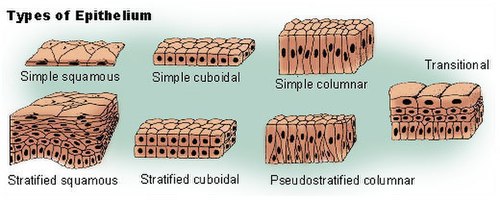
- By layer: simple (single layer), stratified (multiple layers), and pseudostratified (appears layered but all cells contact the basement membrane).
- By cell shape: squamous (flat), cuboidal (cube-like), and columnar (taller than wide).
- Special types: keratinized epithelium of the skin, ciliated epithelia that move particles or fluid, and glandular epithelia that secrete substances such as sweat, oil or mucus (mucus).
- Related linings: endothelium lines blood vessels, and mesothelium lines body cavities—both are specialized epithelial layers serving distinct roles.
Principal functions
Epithelia perform multiple roles depending on location. Key functions include:
- Protection: forming a physical barrier against mechanical injury, chemical exposure and microbial invasion, contributing to innate immunity.
- Selective transport and absorption: moving fluids and solutes into or out of organs, for example intestinal absorption and renal reabsorption (fluid transport).
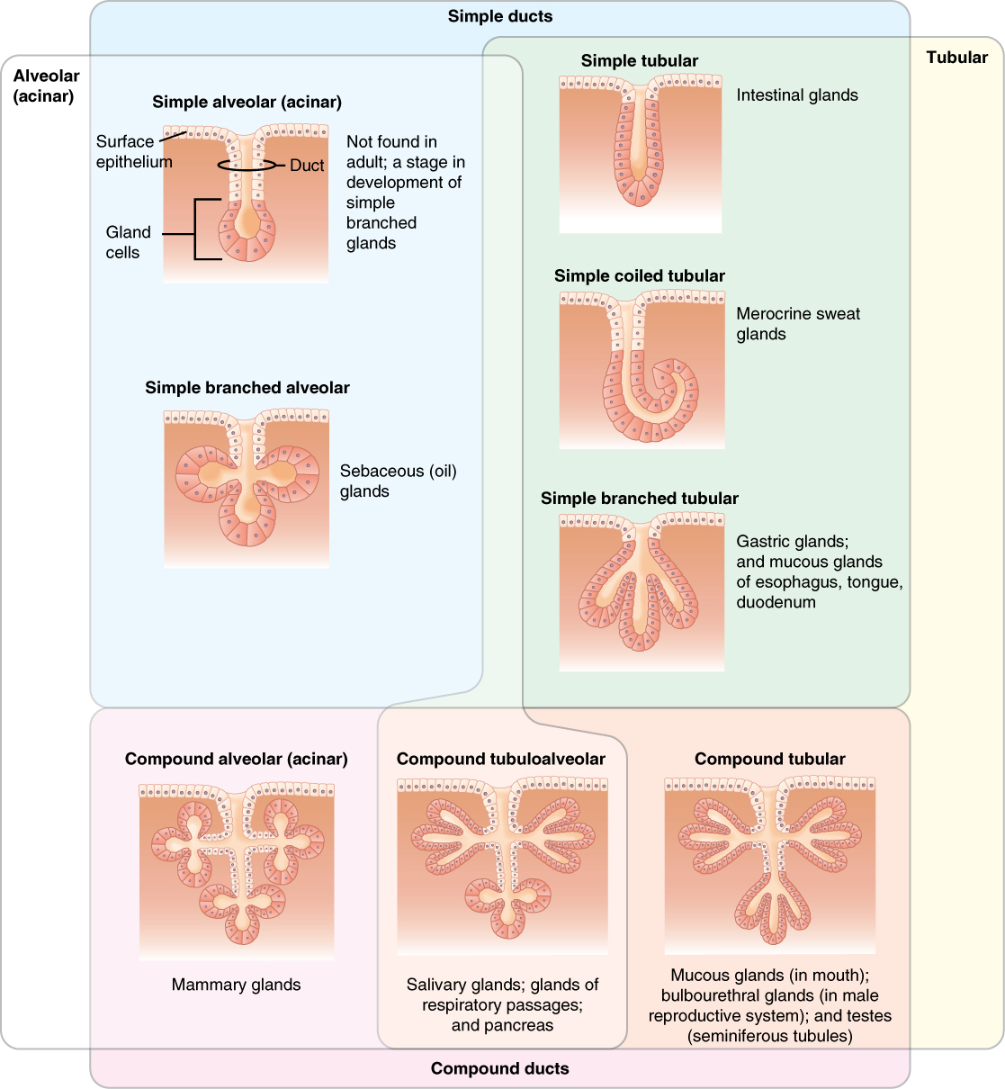
- Secretion: producing enzymes, hormones, mucus and other substances from glandular epithelium; all glands originate from epithelial tissue.
- Sensory roles: specialized epithelial cells detect stimuli in senses such as smell, taste and mechanoreception.
Examples and clinical relevance
Epithelia cover organs such as the stomach and line structures including the kidneys and respiratory tract (organs, stomach). Because epithelial layers are the site of intense interaction with the environment, they are commonly involved in disease: breaches in the barrier can permit infection, chronic irritation can cause metaplasia, and the majority of human cancers that arise from epithelial cells are classified as carcinomas. Disorders of glandular epithelia affect secretion and fluid balance; injuries to the skin epithelium compromise thermoregulation and defence (skin).
Regeneration, diagnosis and research
Many epithelia regenerate rapidly after injury through resident stem cell populations, a property exploited in wound healing and regenerative medicine. Histological examination of epithelial architecture and staining for junctional proteins are routine in diagnostic pathology. Research continues into epithelial barrier function, ciliary motility disorders, and how epithelial signalling influences inflammation and cancer development.
Recognizing epithelial patterns—layering, cell shape, surface specializations and relationship to the basement membrane—helps clinicians and scientists identify tissue type and assess health. For accessible introductions and deeper resources, see referenced summaries and reviews (cells, skin overview, barrier function, innate immunity, organ linings, fluid transport, gastrointestinal epithelium, secretory epithelium, basement membrane and stroma).
Structure
Epithelia are clearly separated from connective tissue by the basement membrane and do not contain blood vessels.
Another property common to all epithelial cells is their polarity:
- The outer, apical side faces the exterior (e.g. in skin) or the lumen (e.g. in intestine or glands).
- The basal side is connected to the underlying tissue by a basal lamina.
The polarity of epithelial cells is also characterized by structural and functional differences between the apical and basal membranes of the epithelial cells. In this context, one also speaks of an apical and basolateral domain.
Furthermore, epithelial cells possess an adhesion complex (junctional complex) consisting of zonula occludens (tight junction), zonula adhaerens (Adhaerens junction) and desmosome (macula adhaerens). On the one hand, the adhesion complex represents a physico-chemical barrier and, on the other hand, connects adjacent epithelial cells with each other.
The cells lie close together and are rich in cell contacts. Consequently, the tissue has only small intercellular spaces with correspondingly little intercellular substance. With the help of emperipolesis, other cells penetrate the epithelia.
Classification of the epithelia
Epithelia are differentiated in many ways and specifically depending on the organ. First of all, one can distinguish surface epithelia and glandular epithelia:
- Surface epithelia primarily have a protective function (e.g. the skin). They can absorb substances (resorption, e.g. intestinal mucosa) and form a barrier that separates the respective organ from the environment (mainly through the already mentioned cell contacts, the tight junctions).
- Glandular epithelia determine the function of all glands (secretion, excretion). They produce secretions of all kinds (among others in salivary glands and sweat glands or in the intestinal mucosa).
For the differentiation of the numerous epithelial types it has proved useful to emphasize two features: First, the number of cell layers and second, the shape of the cells in the superficial cell layer (see below).
Single layer epithelia
Simple epithelia
- Single-layered squamous epithelium: Such epithelia serve primarily as a smooth lining of internal surfaces. Because they are very thin, single-layer squamous epithelia allow for mass transfer (e.g., gas exchange in the alveoli). Examples:
- Endothelium (epithelial lining of blood and lymph vessels)
- Mesothelium (pleural, pericardial, peritoneal epithelium (serous membranes))
- single-layered isoprismatic epithelium (also cubic epithelium): The epithelial cells have an almost cubic shape. These larger cells are metabolically active and perform active transport tasks in terms of secretion/absorption. Examples:
- Kidney tubules
- Glandula submandibularis (salivary glands)
- Bile ducts
- Ovarian Epithelium
- single-layered highly prismatic epithelium ("cylinder epithelium" or also "columnar epithelium"): elongated, columnar cells take over barrier and transport functions with an active metabolism. Examples:
- Gastric mucosa
- Intestinal mucosa
- Fallopian Tube
- Gallbladder
Multiple row epithelia
The multi-rowed epithelium is also still single-layered, all cells are anchored on the basal lamina as in the single-layered epithelium, but not all reach the lumen. Highly prismatic cells perform the actual function, while small basal cells stand by as a reserve for perished cells. The cell nuclei thus lie at different heights, forming apparent layers (rows).
- Ciliated epithelium in the trachea and other airways up to and including the segmental bronchi.
- Vas deferens
- Epididymal ducts
- Eustachian tube
Multilayer epithelia
In the multilayer epithelium, many (more than ten) cell layers lie on top of each other. In principle, a threefold division can be made: Cell divisions take place in the basal layer, which is anchored to the basal lamina. The cells ascend and differentiate in a specific way in a middle or intermediate layer. Finally, they reach the superficial or superficial layer.
- multilayered squamous epithelium: This epithelium is of great importance and is found everywhere where the mechanical load is high. Cytoskeleton and cell contacts are adapted to this load. In regions that are constantly moistened, the multilayer squamous epithelium remains unkeratinized; where it is exposed to air, it keratinizes.
- multilayered unkeratinized squamous epithelium:
- Oral cavity, oesophagus, anal canal
- Vagina
- Cornea and conjunctiva of the eye
- in the male urethra just before the external orifice
- Multi-layered keratinized squamous epithelium: Another protective function is the death and keratinization of the outer cell layers. The cells are massively anchored with desmosomes among each other and with hemidesmosomes in the basal lamina:
- in humans the epidermis is the only keratinizing squamous epithelium
- in ruminants, it is also found in the reticulum, leaf stomach and rumen
- multilayer isoprismatic epithelium: ovarian follicles that have reached the stage of secondary follicle have such an epithelium.
- bilayered isoprismatic epithelium: This type of epithelium is found in the excretory ducts of the sweat glands. The ciliary body is also covered by such an epithelium, but it is part of the retina.
- multilayered high prismatic epithelium: This less common form of epithelium is to be distinguished from the much more significant multilayered high prismatic epithelium. It is found in only three locations in the human body:
- in the male urethra in its course from the prostate to just before the external orifice
- in main excretory ducts of the large salivary glands (two-layered)
- in the fornix conjunctivae, a reserve fold of the conjunctiva
Transitional epithelium ("urothelium")
The transitional epithelium ("urothelium") is a special epithelium of the urinary tract (renal pelvis, ureter, urinary bladder) with several rows to several layers, depending on the filling of the bladder (or stretching of the urothelium). Here, the covering/umbrella cells are of particular importance. They form the so-called crusta, which has the task of uric acid protection. In contrast to the squamous epithelium, the upper cell layer is more cubic.


Related articles
Author
AlegsaOnline.com Epithelium: structure, types, functions and clinical significance Leandro Alegsa
URL: https://en.alegsaonline.com/art/31766
Sources
- books.google.com : Dellmann's textbook of veterinary histology
- books.google.com : p. 3
- britannica.com : Article fromEncyclopedia Britannica regarding Epithelium