Venule: small vessels of the microcirculation
A venule is a small vessel that collects blood from capillary beds and delivers it to veins. Venules play key roles in microcirculation, fluid balance, and immune cell trafficking.
Overview: A venule is a very small blood vessel that collects blood from the capillary network and channels it toward larger veins. Venules bridge the microcirculation between capillaries and veins and are essential for returning deoxygenated blood to the heart. For general context see blood vessel and the movement of blood. They form where individual capillaries converge and ultimately drain into larger veins. Typical diameters range broadly (commonly cited between 8 and 100 µm) depending on location and function (size reference).
Image gallery
1 Image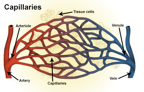
Structure and microscopic features
Venules are lined by a single layer of endothelial cells resting on a basement membrane. Unlike arterioles, the tunica media is minimal or absent in the smallest venules; pericytes and scattered smooth muscle cells appear as venules enlarge. The wall layers are thinner and more distensible than those of arteries, which helps venules accommodate changes in blood volume and pressure within the microcirculation.
Function and physiological roles
Primary functions of venules include collecting blood from capillaries and acting as a low-resistance conduit toward veins. Postcapillary venules are especially important for regulating tissue fluid balance and for immune surveillance: they are the principal sites where white blood cells leave the circulation (diapedesis) during inflammation. Venular endothelium also modulates permeability under the influence of mediators such as histamine, promoting transient fluid and plasma protein exit into tissues when required.
Types and distinguishing features
- Postcapillary venules: smallest venules directly downstream from capillaries; major sites of leukocyte extravasation.
- Collecting venules: larger, with more defined supporting cells and some smooth muscle.
- Muscular venules: have several layers of smooth muscle and act more like small veins.
- High endothelial venules (HEVs): specialized venules in lymphoid tissue with cuboidal endothelium that supports lymphocyte trafficking.
Development, relationships and distinctions
Venules develop by the union of capillary channels and by remodeling during angiogenesis. In the microcirculation they occupy the low-pressure, high-capacitance side, contrasting with arterioles that regulate resistance and perfusion. Venules generally lack the robust muscular wall and internal elastic lamina found in arterioles and larger arteries.
Clinical relevance
Altered venular function contributes to edema when permeability or hydrostatic pressure increases, and to inflammation when leukocyte recruitment is excessive. Conditions described as venulitis involve inflammation of venules; although thrombosis is less common in small venules than in larger veins, microvascular obstruction can occur in severe infections or vasculopathies. Understanding venule biology is important for therapies targeting inflammation, vascular permeability, and immune cell trafficking.
Related articles
Author
AlegsaOnline.com Venule: small vessels of the microcirculation Leandro Alegsa
URL: https://en.alegsaonline.com/art/104599