Sublingual gland
The sublingual glands are the smallest of the major salivary glands, located beneath the tongue; they produce mainly mucous saliva and have several small ducts that open into the floor of the mouth.
Overview
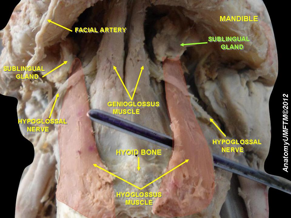
The sublingual glands are paired salivary glands situated beneath the oral mucosa on the floor of the mouth, immediately under the tongue. They are classed with the major salivary glands alongside the parotid and submandibular glands. Their secretions contribute to lubrication, digestion, and protection of the oral cavity. For general context about salivary glands see related overview.
Image gallery
8 Images


Structure and anatomy
Each sublingual gland is small and almond-shaped, lying anterior to the submandibular gland and medial to the mandible. Unlike the larger glands, the sublingual has multiple small excretory ducts, often called the ducts of Rivinus, which open along the sublingual fold on the floor of the mouth. A larger duct, the duct of Bartholin, sometimes joins the submandibular (Wharton) duct before reaching the oral cavity. Anatomical details are discussed in many clinical references: anatomy source.
Secretion and innervation
The sublingual glands secrete primarily mucous saliva, producing a viscous fluid that helps form the mucous layer of saliva. Parasympathetic fibers stimulate secretion; these travel from the facial nerve via the chorda tympani to the submandibular ganglion. Sympathetic fibers modulate blood flow and composition of the saliva. Functional summaries and physiology can be found at physiology notes.
Clinical relevance
- Ranula: mucus retention cysts may arise from obstruction of a sublingual duct and present as a swelling in the floor of the mouth.
- Sialolithiasis: salivary stones are less common here than in the submandibular gland but can occur and cause pain or infection.
- Inflammation and tumors: infections or neoplasms are relatively uncommon but clinically important when they occur.
Surgical removal (sublingual sialadenectomy) or marsupialization of a ranula are among the treatments; typical management strategies are described in clinical guidelines: clinical guidance.
Notable facts and distinctions
The sublingual glands are the smallest of the major glands and differ from the parotid (mostly serous secretion) and submandibular glands (mixed secretion) by their predominantly mucous output and multiplicity of ducts. Their location beneath the tongue makes them accessible in intraoral examinations and important to consider in procedures involving the mouth floor and tongue base.
Related articles
Author
AlegsaOnline.com Sublingual gland Leandro Alegsa
URL: https://en.alegsaonline.com/art/94491