
Heart valve: structure, function, disorders and treatments
A heart valve regulates one-way blood flow through the heart. This article covers the main valves, their anatomy and mechanics, common diseases, diagnosis, and treatment approaches.
Overview
Heart valves are thin, fibrous structures that direct the circulation of blood through the chambers of the heart. Each valve normally permits flow in one direction and prevents backward flow, using changes in blood pressure across the valve to open and close. Proper valve function is essential for efficient pumping and for maintaining the separation between high‑pressure systemic and lower‑pressure pulmonary circuits.
Image gallery
6 Images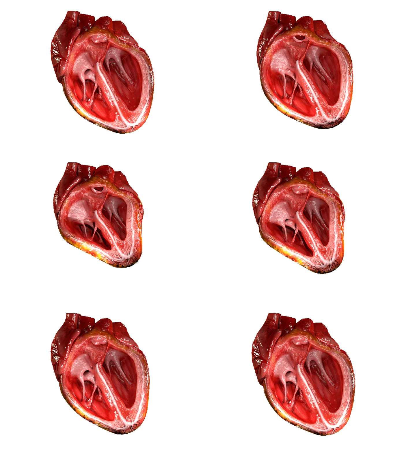



Anatomy and types
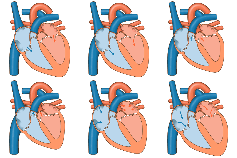
Mammals typically have four principal valves organized into two categories. The atrioventricular (AV) valves separate atria from ventricles, and the semilunar valves separate ventricles from the arterial outflow tracts. The four valves are:
- Tricuspid valve — between right atrium and right ventricle.
- Mitral (bicuspid) valve — between left atrium and left ventricle.
- Pulmonary valve — at the exit of the right ventricle into the pulmonary artery.
- Aortic valve — at the exit of the left ventricle into the aorta.
AV valves are supported by fibrous rings called annuli and, on the ventricular side, by chordae tendineae and papillary muscles that prevent prolapse during ventricular contraction. Semilunar valves have three pocket‑shaped cusps and lack chordae.
Function and mechanics
Valves respond passively to differences in blood pressure across their leaflets. When upstream pressure exceeds downstream pressure, the valve opens to allow blood flow. When downstream pressure rises above upstream pressure, the leaflets coapt and close, preventing regurgitation. Effective closure and leaflet mobility depend on leaflet tissue, the geometry of the annulus, and coordinated contraction of supporting muscles.
Clinical significance
Valve disease can be congenital or acquired. Common problems include stenosis (narrowing that obstructs outflow), regurgitation (leakage backward), and infectious or inflammatory injury (endocarditis, rheumatic damage). Symptoms vary with severity and can include breathlessness, fatigue, palpitations, and heart murmurs detected on auscultation.
Diagnosis and treatment
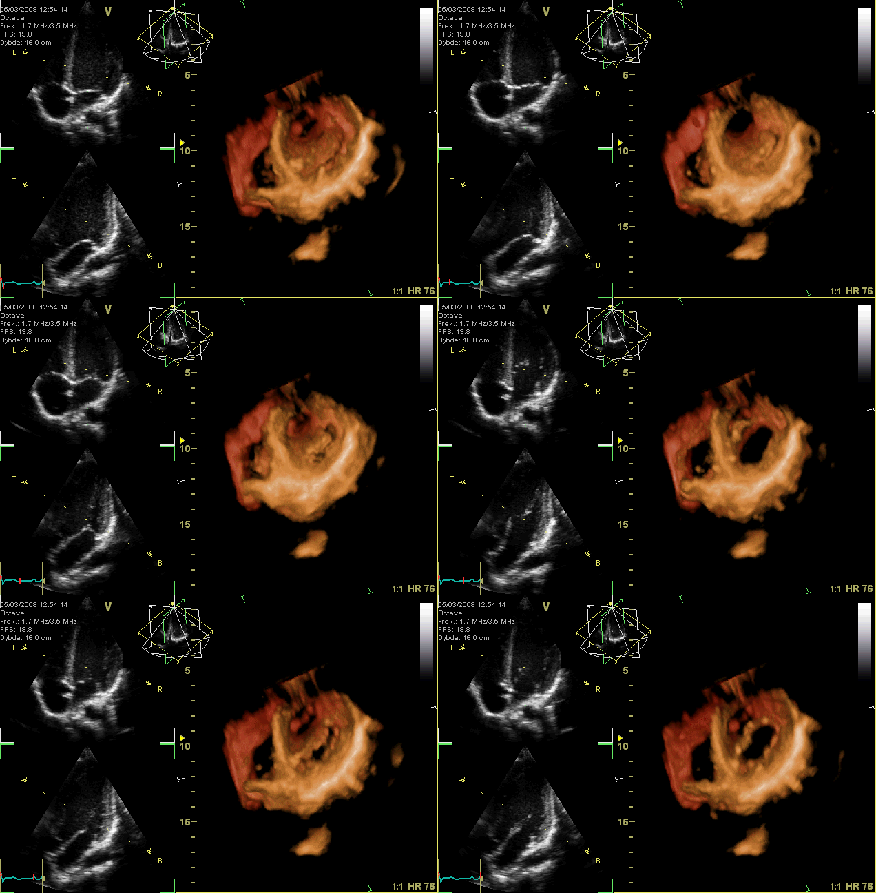
Evaluation typically uses physical examination and imaging such as echocardiography to visualize structure and measure severity. Management ranges from medical monitoring to interventional procedures. Typical approaches include:
- Conservative care and medications for symptom control.
- Repair techniques that restore native valve function.
- Valve replacement with mechanical or tissue prostheses.
- Transcatheter interventions (less invasive valve implantation or balloon dilation) for select patients.
Because valves control directional blood flow and influence cardiac workload, preserving or restoring valve competence is a central goal in treating many forms of heart disease.
Historical and notable facts
The role of valves in circulation became clear as knowledge of cardiac physiology developed; understanding their mechanics was fundamental to later advances in cardiac surgery and prosthetic design. Modern valve care combines imaging, surgical technique, and catheter-based technologies to reduce symptoms and improve survival in patients with significant valve disease.
Related articles
Author
AlegsaOnline.com Heart valve: structure, function, disorders and treatments Leandro Alegsa
URL: https://en.alegsaonline.com/art/43090
Sources
- americanheart.org : "Heart Valves"
- cvphysiology.com : "Pressure Gradients"
- cvphysiology.com : "Cardiac Valve Disease"