Aneurysm: causes, types, diagnosis and treatment
An aneurysm is a localized dilation of a blood vessel due to weakened vessel wall. This article explains types, common locations, causes, symptoms, diagnosis, treatment and prevention.
An aneurysm is a focal enlargement of a blood-filled part of a vessel caused by weakness in the vessel wall. Left untreated, an aneurysm may grow and eventually rupture, producing severe bleeding and life-threatening complications. The term covers a range of lesions that differ by shape, cause and location.
Image gallery
6 Images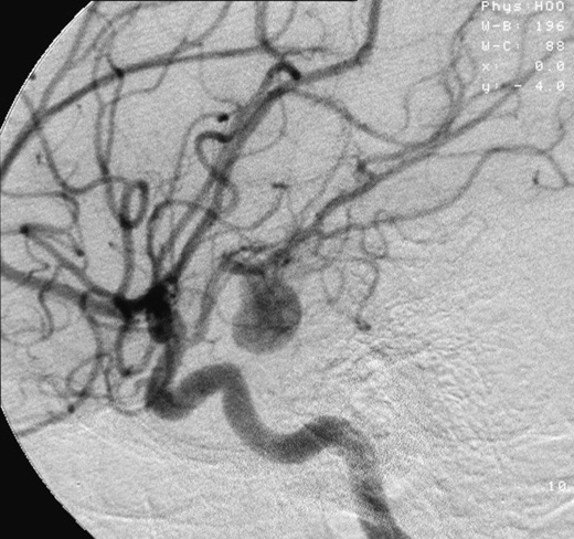


Types and structural distinctions
Aneurysms are described by their shape and by whether all layers of the vessel wall are involved. Common categories include:
- Saccular (berry) — a round outpouching with a narrow neck.
- Fusiform — a more uniform, spindle-shaped dilatation affecting the entire circumference.
- True vs pseudoaneurysm — true aneurysms involve the intact vessel wall layers; pseudoaneurysms are contained ruptures where blood is held by surrounding tissue.
Common locations and clinical importance
Aneurysms most often affect large arteries exposed to high pressure. Two clinically important sites are cerebral arteries at the base of the brain and the aorta leaving the heart. Intracranial aneurysms can cause subarachnoid hemorrhage if they rupture. Aortic aneurysms, whether thoracic or abdominal, can result in massive internal bleeding.
Key vascular and anatomical terms are often linked to further discussion: blood, blood vessel, disease, arteries, brain, heart and aortic aneurysm.
Causes and risk factors
Wall weakness has multiple contributors. Chronic high blood pressure and atherosclerosis weaken vessels over time. Smoking, advancing age, infection or trauma and certain inherited connective tissue disorders (for example, Marfan or Ehlers–Danlos syndromes) increase risk. Hemodynamic stress at arterial branch points also predisposes to sac-like intracranial aneurysms.
Symptoms, diagnosis and treatment
Many aneurysms produce no symptoms until they enlarge or rupture. Unruptured aneurysms may cause local pressure effects — headaches, focal neurological signs, or pain — depending on location. Rupture leads to sudden severe headache, loss of consciousness, shock, or death.
Diagnosis uses imaging appropriate to the suspected site: ultrasound for abdominal aortic aneurysm, CT or MRI for brain lesions, and catheter angiography or CT angiography for detailed vascular mapping. Management ranges from watchful surveillance and blood pressure control to procedural repair. Options include endovascular techniques (coiling, stent graft placement) and open surgical repair (clipping of cerebral aneurysms, graft replacement of aortic segments).
Prevention focuses on controlling blood pressure, stopping smoking, and selective screening of high-risk groups. Advances in endovascular devices since the late 20th century have broadened treatment choices and reduced recovery time for many patients, but decision-making remains individualized and depends on aneurysm size, location and patient risk.
Related articles
Author
AlegsaOnline.com Aneurysm: causes, types, diagnosis and treatment Leandro Alegsa
URL: https://en.alegsaonline.com/art/4095