
Urinary bladder: structure, function, control, and clinical significance
The urinary bladder is a hollow, muscular organ that stores urine and coordinates its controlled release. This article covers anatomy, physiology, neural control, development, and common disorders.
Overview
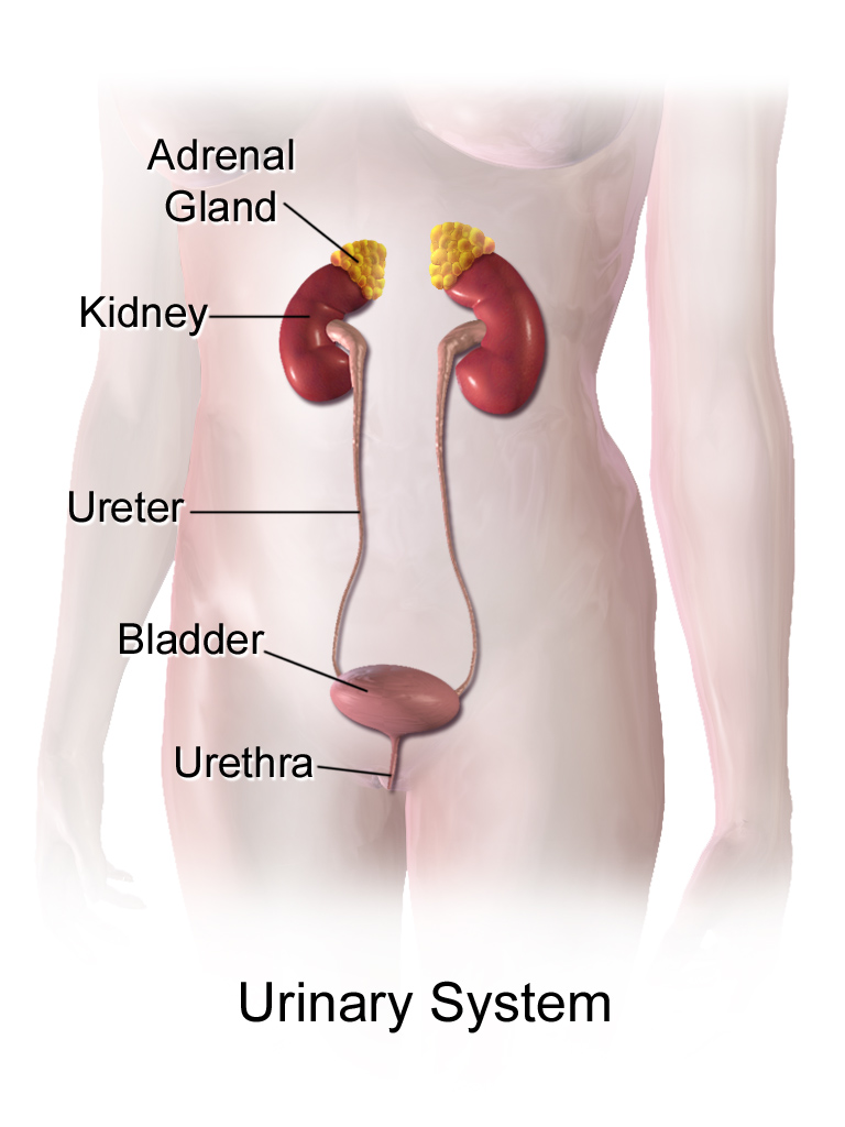
The urinary bladder is a hollow, distensible organ in the human body that temporarily stores urine before voluntary or reflex elimination. It is a central component of the urinary system, receiving urine produced and concentrated by the kidneys and passing it to the urethra. The bladder allows elimination to be intermittent rather than continuous, helping maintain fluid balance and protect the upper urinary tract.
Image gallery
10 Images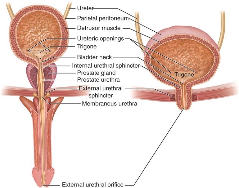





Structure and layers
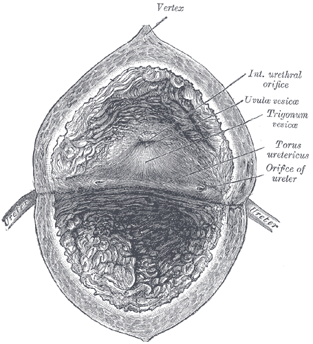
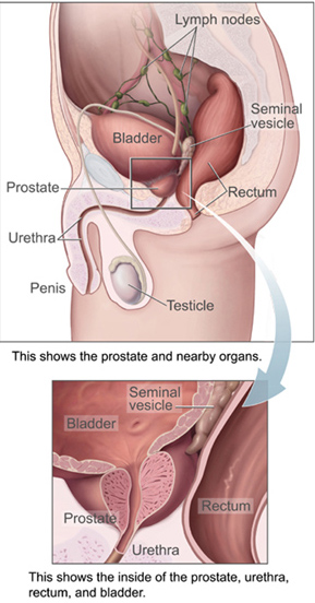
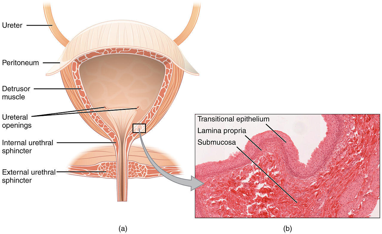
Anatomically the bladder is a muscular sac with several distinguishable regions. The inner lining is a specialized epithelium called the urothelium that forms a barrier to urine. Beneath the urothelium is connective tissue and then the detrusor muscle, a layer of smooth muscle responsible for contraction. The bladder neck and urethral sphincters provide outflow control. In men the bladder lies above the prostate; in women it is positioned anterior to the uterus and vagina, which contributes to some differences in clinical presentation.
Function and neural control
The bladder's primary role is storage and controlled emptying. It receives liquid from the ureters and expands; the urothelium and sensory nerves signal filling to the central nervous system. When appropriate, parasympathetic pathways trigger detrusor contraction while sympathetic and somatic pathways relax internal and external sphincters. This coordinated sequence—often centered in spinal and brainstem circuits, including the pontine micturition center—produces the act of voiding. The bladder works with other tissues and signals: it senses changes in volume and composition of fluids, communicates with the brain, and is effected by autonomic and voluntary muscles that can contract or relax.
Development and life stages
The bladder develops from the embryonic urogenital sinus and matures over childhood; control of urinary storage and voiding is learned as neural pathways and pelvic muscles develop. Age-related changes may alter capacity, sensation, and contractility, and conditions such as pregnancy or prostate enlargement can modify bladder function.
Clinical importance and common conditions
- Infections (cystitis) and stones that irritate the bladder lining.
- Urinary incontinence from sphincter weakness, pelvic floor dysfunction, or neurologic disease.
- Urinary retention caused by obstruction or poor detrusor contractility.
- Neurogenic bladder resulting from spinal cord injury, stroke, or neuropathy.
- Bladder cancer arising from the urothelium, often detected by hematuria or imaging.
Diagnostic tools include urinalysis, ultrasound, cystoscopy, and urodynamic testing. Treatments range from behavioral therapies and pelvic rehabilitation to medication, catheterization, and surgery.
Notable distinctions
Unlike many hollow organs, the bladder can change shape and volume dramatically without large changes in internal pressure, due to its compliant wall and specialized lining. Its function depends on precise timing between sensory signals and motor responses; disruption of any element—epithelium, muscle, nerves, or outlet—can produce symptoms. Understanding bladder biology is important for managing common conditions that affect quality of life and urinary tract health.
Further reading: anatomical atlases and clinical urology texts provide detailed images and protocols—consult specialist resources for diagnostic or treatment decisions.
More on bladder anatomy | Human anatomy overview | Urinary system | Body fluids | Kidneys and urine formation | Neural control of micturition | Pelvic muscles | Detrusor function
Related articles
Author
AlegsaOnline.com Urinary bladder: structure, function, control, and clinical significance Leandro Alegsa
URL: https://en.alegsaonline.com/art/12039