
Rete testis: anatomy, function and clinical significance
The rete testis is an intratesticular network of channels that conveys sperm from seminiferous tubules to efferent ductules. This article explains its structure, role, development and common clinical issues.
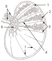
The rete testis is an intratesticular network of thin channels located in the mediastinum of the testis that collects spermatozoa and testicular fluid produced by the seminiferous tubules. It forms the first conduit in the male reproductive tract inside the testis, receiving sperm from the straight tubules and directing them to the efferent ductules and the epididymis. Anatomically it sits near the testicular hilum and is best understood as a bridging system rather than a single continuous tube.
Image gallery
6 Images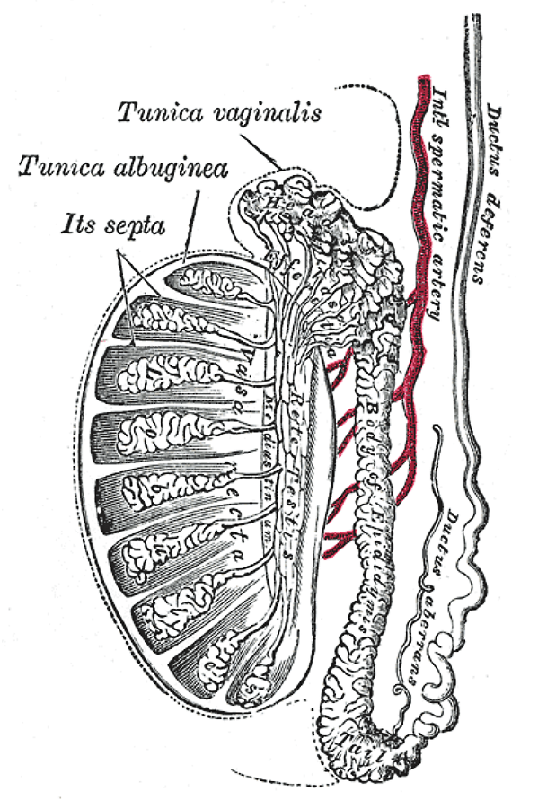


Structure and histology
Microscopically, the rete testis consists of interconnected tubules lined by simple cuboidal or low columnar epithelium. The cells may show microvilli and tight junctions that help regulate the composition of luminal fluid. The network is embedded within connective tissue of the mediastinum testis and contains a mixture of spermatozoa and fluid that is moved by pressure gradients and subtle contractions from surrounding tissue.
Development and relationships
During fetal development the rete testis forms as the intratesticular channels that link seminiferous tubules to the efferent ductules. Those efferent ductules connect externally to the epididymis. For a general overview of testicular anatomy, consult testis anatomy. For a focused description of the rete itself see Rete testis.
Function and clinical importance
- Function: transports sperm and participates in modification of tubular fluid prior to epididymal maturation.
- Common clinical issues: benign cysts of the rete testis are detectable on ultrasound and are usually harmless; information on such lesions is often listed under rete cysts.
- Other concerns: obstruction, inflammation or very rare tumors can affect fertility or require further investigation.
Imaging with scrotal ultrasound is the usual method to evaluate suspected rete abnormalities, showing small anechoic or hypoechoic spaces near the mediastinum. When benign features are clear, conservative management is typical; atypical findings prompt urologic assessment. Distinguishing rete abnormalities from epididymal or paratesticular lesions relies on location, ultrasound pattern, and clinical context.
Related articles
Author
AlegsaOnline.com Rete testis: anatomy, function and clinical significance Leandro Alegsa
URL: https://en.alegsaonline.com/art/82336