
Radiology: Medical Imaging, Methods, History, and Applications
Radiology is the medical specialty that produces and interprets images of the body to diagnose and treat disease, using modalities such as X‑ray, CT, MRI, ultrasound and PET.
Overview: Radiology is the medical discipline that uses imaging technologies to visualize internal structures and functions for diagnosis, monitoring and treatment. Radiologists work alongside clinicians to interpret images, guide procedures, and advise on the safest, most effective imaging strategy for each patient. Imaging contributes to most areas of modern healthcare, from emergency care to chronic disease management.
Image gallery
5 Images


Common imaging modalities
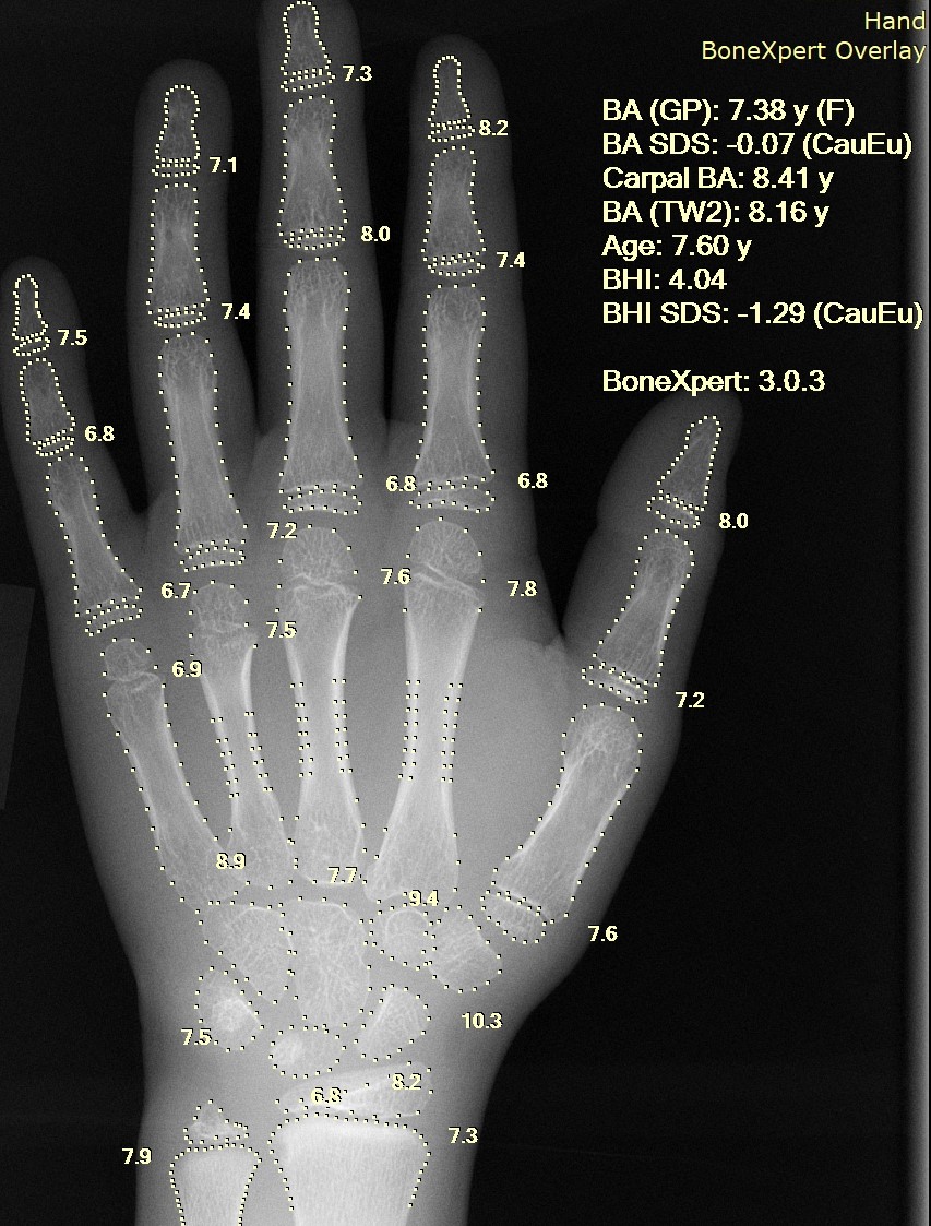
- X‑ray (radiography) — fast, widely available projection images useful for bones, chest and many acute conditions.
- Ultrasound (sonography) — uses sound waves for real‑time imaging of soft tissues, obstetrics, and vascular flow.
- Computed tomography (CT) — cross‑sectional X‑ray scans that show detailed anatomy and are essential in trauma and cancer staging.
- Positron emission tomography (PET) — a functional technique that maps metabolic activity, often combined with CT for oncologic assessment.
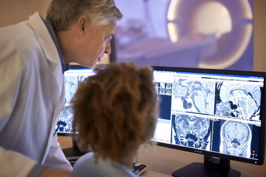
- Magnetic resonance imaging (MRI) — uses magnetic fields and radio waves to produce high‑contrast images of soft tissues and the nervous system.
Principles and distinctions: Modalities differ by physical principle (ionizing radiation vs non‑ionizing), by whether they emphasize anatomy or function, and by their resolution and speed. Radiography and CT rely on X‑rays; CT provides cross‑sectional detail while radiographs are two‑dimensional. MRI and ultrasound avoid ionizing radiation; MRI excels at soft tissue contrast. PET and some MRI techniques provide physiological or molecular information rather than only structure.
History and development: Modern radiology began after Wilhelm Röntgen's discovery of X‑rays in 1895. Cross‑sectional CT emerged in the late 20th century, transforming diagnostic capability, while MRI and PET added advanced soft‑tissue and metabolic imaging. Over decades, improvements in detectors, computing and contrast agents have steadily increased image quality and clinical utility.
Clinical uses and procedures: Radiology supports diagnosis (fractures, infections, tumors), guides interventions (angiography, biopsies, drainages), and monitors therapy response. Interventional radiology uses imaging guidance to perform minimally invasive treatments such as vascular stenting, embolization and image‑guided ablation, reducing the need for open surgery.
Training, safety and future directions: Radiologists undergo specialist training and collaborate with technologists and medical physicists to optimize protocols and minimize risk. Radiation safety, appropriate use criteria and contrast‑related precautions are central concerns. Emerging trends include hybrid scanners (PET/CT, PET/MRI), quantitative imaging, and artificial intelligence tools that assist interpretation and workflow, all of which aim to improve accuracy and personalize care.
For further general information about the role of imaging in clinical care see clinical resources and diagnostic guidelines at authoritative sources on recommendations or modality summaries for ultrasound, for CT and for PET.
Related articles
Author
AlegsaOnline.com Radiology: Medical Imaging, Methods, History, and Applications Leandro Alegsa
URL: https://en.alegsaonline.com/art/80777