
Positron Emission Tomography (PET): Principles, Uses, and Limitations
Positron emission tomography (PET) is a medical imaging method that visualizes physiological processes using short‑lived radioactive tracers. Common applications include cancer detection, brain studies, and cardiac assessment.
Overview
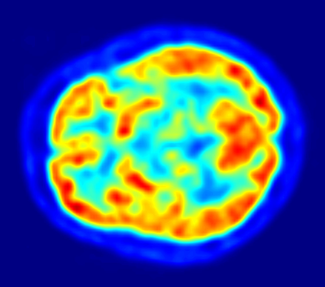
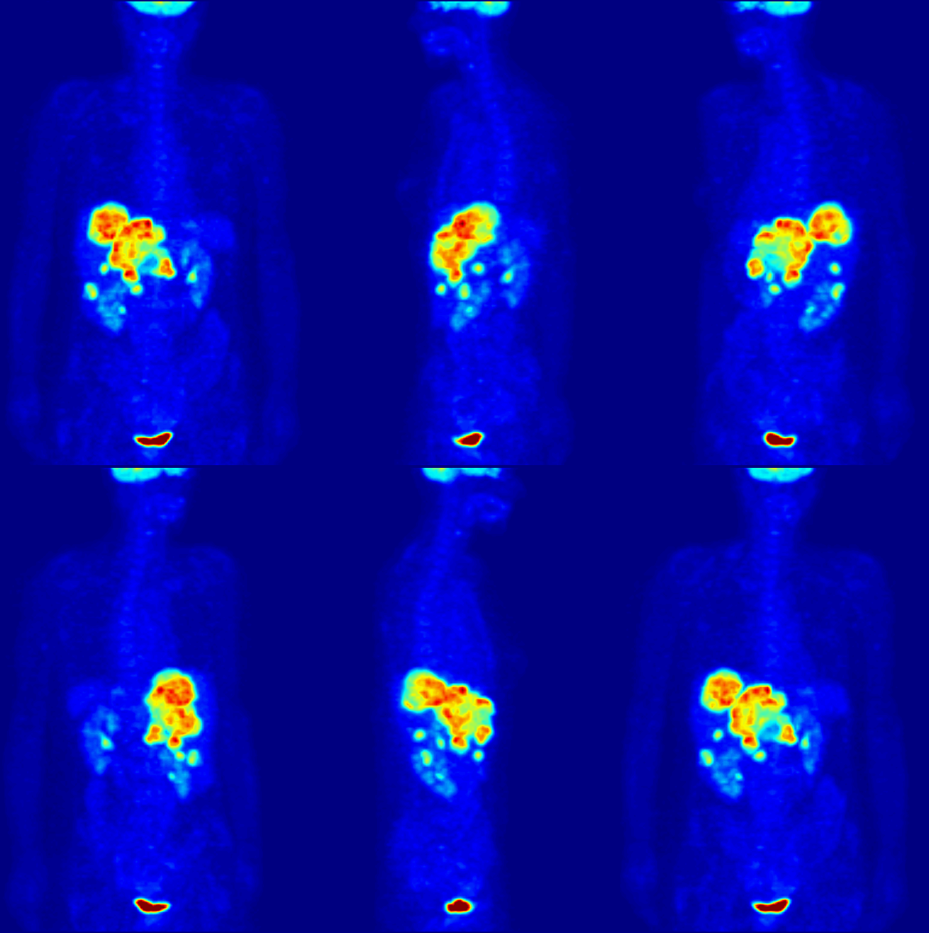
Positron emission tomography (PET) is a functional imaging technique used in medicine and biomedical research to visualize biochemical and physiological processes in the body. Instead of showing anatomy like X‑ray or MRI, PET maps the distribution of a radiolabeled substance — commonly called a tracer — which accumulates where specific biological activity occurs. Data from a PET scan are rendered as colorized images that highlight regions of higher or lower tracer uptake, enabling clinicians and researchers to infer metabolic, receptor, or perfusion patterns.
Image gallery
10 Images





How PET works
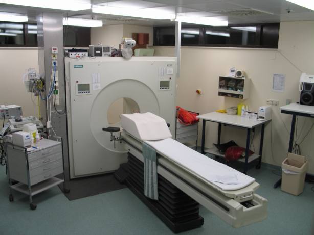
A PET study begins with administration of a tracer that emits positrons as it decays. When a positron encounters an electron in tissue, the two annihilate and produce a pair of gamma photons that travel in nearly opposite directions. A ring of detectors surrounding the patient records coincident photon events; software reconstructs these events into a three‑dimensional image showing where annihilations — and thus tracer concentration — occurred. Modern systems use scintillation crystals and coincidence timing to improve sensitivity and spatial resolution, and images are often corrected for attenuation and scatter to improve accuracy.
Tracers and production
Tracers vary according to the physiological process of interest. The most widely used tracer in clinical PET is a glucose analogue labelled with the radioisotope fluorine‑18, which highlights regions of high glucose metabolism such as many tumors or active brain areas. Other tracers target neurotransmitter receptors, amyloid plaques in neurodegenerative disease, or myocardial blood flow. Because the radioactive isotopes used decay quickly, tracers must be produced close to the imaging site, typically in a cyclotron and synthesized in a radiochemistry laboratory. That short radioactive half-life and the need for specialized production facilities affect logistics and cost. The administered substance itself is often referred to as a tracer in clinical reports and research papers.
Clinical and research applications
- Oncology: staging, detecting recurrence, and monitoring response to therapy.
- Neurology: evaluating epilepsy, dementia, movement disorders, and brain metabolism.
- Cardiology: assessing myocardial viability and perfusion.
- Drug development and basic research: measuring pharmacokinetics, receptor occupancy, and molecular pathways.
Advantages, limitations and notable facts
PET provides sensitive, quantitative information about function that complements anatomical imaging. Combined systems such as PET/CT and PET/MRI fuse metabolic data with high‑resolution anatomy, improving lesion localization and diagnostic confidence. Limitations include limited spatial resolution compared with structural imaging, exposure to ionizing radiation, high equipment and operational costs, and dependency on short‑lived tracers that require nearby production. Despite these constraints, PET remains a cornerstone of precision diagnosis, treatment planning, and biomedical research because it can reveal disease activity at a molecular level before structural changes appear.
Practical considerations
Preparation and interpretation of PET studies involve multidisciplinary teams: nuclear medicine physicians, radiochemists, technologists, medical physicists and referring clinicians. Patient preparation often includes fasting or other protocol‑specific instructions to optimize tracer distribution. As tracer chemistry and detector technology evolve, PET continues to expand its role across clinical specialties and translational research.
Questions and answers
Q: What is Positron Emission Tomography (PET)?
A: PET is a medical imaging technique used to show the path and distribution of a weakly radioactive substance.
Q: What is the tracer used in PET scans?
A: The tracer used in PET scans is a weakly radioactive substance that dissolves in the blood and concentrates around active brain structures.
Q: What is the purpose of PET scans?
A: The purpose of PET scans is to detect tumors and highlight active areas of the brain.
Q: How is the radioactivity detected in PET scans?
A: The radioactivity in PET scans is detected by a specially constructed device which converts it into a digital image of the brain.
Q: What kind of tracers are used in PET scans?
A: Different kinds of tracers are used in PET scans, but they are often similar to sugar or specially modified sugars.
Q: What is the half-life of the tracer used in PET scans?
A: The half-life of the tracer used in PET scans is around 30-60 minutes.
Q: Why is producing the tracer difficult and expensive?
A: Producing the tracer is difficult and expensive due to the different factors involved in preparing the tracer for the event as it cannot be mass-produced.
Related articles
Author
AlegsaOnline.com Positron Emission Tomography (PET): Principles, Uses, and Limitations Leandro Alegsa
URL: https://en.alegsaonline.com/art/78347