Magnetic Resonance Imaging (MRI): Principles, Technology and Clinical Uses
Comprehensive overview of MRI: physical principles, system components, common clinical applications, historical milestones, and safety considerations for this radiation-free imaging method.
Overview
Magnetic resonance imaging (MRI) is a medical imaging technique used to produce detailed pictures of the body's internal soft tissues, organs, and structures, including muscle and other flesh. Unlike X-rays or CT scans, MRI does not rely on ionizing radiation. It exploits the physics of nuclear magnetic resonance, a phenomenon in which certain atomic nuclei respond to applied magnetic and radiofrequency fields.
Image gallery
10 Images


How MRI works
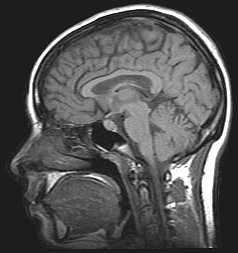
In an MRI examination the patient lies on a moveable table that slides into the scanner bore. A strong main magnetic field aligns the magnetic moments (spins) of hydrogen nuclei in the body. Short bursts of radio waves are then transmitted to perturb that alignment. When the radiofrequency pulse ends, the nuclei relax back toward equilibrium and emit weak electromagnetic signals. Those signals contain spatial and tissue-contrast information that the scanner receives and forwards to a processing unit.
Main components of an MRI system
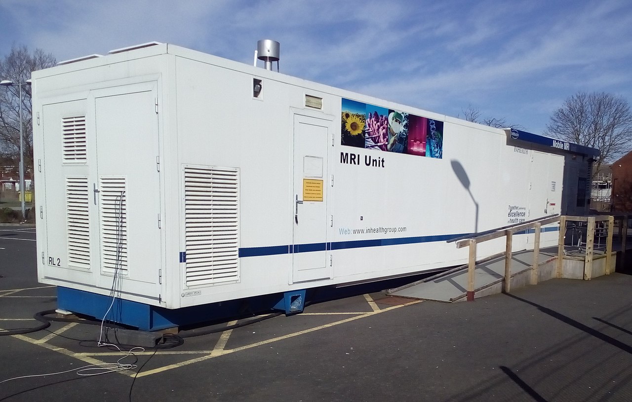
An MRI scanner consists of several principal parts. The superconducting or permanent magnet provides the stable main field; gradient coils impose controlled variations in the field for spatial encoding; radiofrequency (RF) coils transmit pulses and receive the resulting signals; and the central computer reconstructs images. The physical device that houses these elements is commonly called the MRI scanner.
Image formation and common sequences
Images arise after applying combinations of RF pulses and gradients and then digitizing the emitted signals. Fourier transform techniques convert the raw data into cross-sectional images. Different pulse sequences emphasize distinct tissue properties: for example, T1-weighted images provide anatomical detail, T2-weighted images highlight fluid and edema, and diffusion-weighted imaging detects restricted water movement useful in stroke assessment. Magnetic resonance angiography and spectroscopy are additional specialized methods.
Clinical applications and examples
- Neurology: brain tumors, multiple sclerosis, stroke evaluation, and functional MRI mapping of brain activity.
- Musculoskeletal: joint injuries, cartilage assessment, and soft-tissue tumors.
- Cardiology: structural heart disease, myocardial viability, and flow measurements.
- Body imaging: liver, kidneys, pelvis, and vascular studies without ionizing radiation.
History, comparisons and safety
Developed from mid-20th-century discoveries in nuclear magnetic resonance, practical imaging systems appeared in the 1970s and matured into a routine clinical tool by the 1980s. MRI offers superior soft-tissue contrast compared with CT, but it is typically slower, more expensive, and sensitive to patient motion. Strong magnetic fields mean certain metallic implants and devices are contraindicated; patient screening is essential. Gadolinium-based contrast agents are used selectively to improve lesion detection, though their use follows safety guidelines. For more technical or patient-focused resources, see additional materials at technical overview and device information at system specifications or physical principles.
Notable facts: Functional MRI (fMRI) maps brain activity indirectly via blood oxygenation changes; typical clinical field strengths include 1.5 and 3 tesla; and rapid developments continue in areas such as faster sequences, higher-field scanners, and hybrid imaging.
For general background and patient guidance, consult manufacturer and institutional resources available through device documentation and clinical radiology departments: patient information, scanner manuals, and research articles accessible from academic repositories at research portals.


Methods and systems
Numerous special MRI techniques have been developed to provide not only the position and shape of organs but also information about their microstructure and function (especially their blood flow), for example:
- real-time MRI for the cinematic representation of moving joints or organs (e.g. heart),
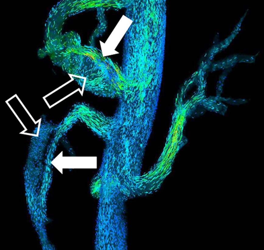
- magnetic resonance angiography (MRA) for imaging the vessels,
- functional magnetic resonance imaging (fMRI) of the brain,
- perfusion MRI for the examination of tissue perfusion,
- diffusion or diffusion tensor imaging (DTI) for a virtual reconstruction of nerve fiber connections,
- MR elastography.
A distinction is made between closed MRI systems with a short or long tunnel and open MRI systems (oMRI) with a C-arm or laterally open tunnel. Closed tunnel systems provide comparatively better image data, whereas open MRI systems allow access to the patient under MRI control.
Another distinguishing criterion is the type of magnetic field generation. For weak magnetic fields up to approx. 0.5 Tesla flux density (magnetic induction), permanent magnets or conventional electromagnets are used, whereas superconducting magnetic coils are used for higher fields.
Image Review
The signal strength of the voxels is represented in coded gray values. Since it depends on numerous parameters (such as the magnetic field strength), there are no standard values for the signal of certain tissues and no defined unit, comparable to the Hounsfield units in computer tomography. The MR console displays only arbitrary (arbitrary) units that are not directly useful diagnostically. Instead, image interpretation relies on the overall contrast, the respective weighting (synonymously weighting) of the measurement sequence, and the signal differences between known and unknown tissues. Therefore, when describing a lesion in the findings, we do not speak of "light" or "dark", but of hyperintens for signal-rich, light and of hypointens for signal-poor, dark.
Depending on the weighting, the different tissues are displayed in a characteristic intensity distribution:
- In T1 weighting, fatty tissue appears hyperintense (signal-rich, bright) and thus also fatty/rich tissue (e.g. bone marrow). This weighting is therefore well suited for the anatomical depiction of organ structures and, especially after contrast medium administration (gadolinium), for better delineation of unknown structures (e.g. tumor).
- In the T2 weighting, stationary fluids appear hyperintense, so that fluid-filled structures (e.g. cerebrospinal fluid spaces) appear signal-rich and thus bright. This makes this weighting suitable for visualizing effusion and edema and, for example, for differentiating cysts from solid tumors. In contrast, in X-ray images, especially in the special X-ray technique of computed tomography (CT), the terms hyperdens and hypodens are used to describe the relative degree of darkness.
- Proton weighted images are faint but sharp. Cartilage can be assessed in great detail. In combination with a fat saturation pulse, PD images are therefore standard in joint examinations.
In voxel-based morphometry, MR images are processed algorithmically to determine objective parameters and analyze them statistically. These methods are used in particular to determine the size of certain brain structures when examining the human brain.
Questions and answers
Q: What is MRI?
A: MRI stands for Magnetic Resonance Imaging, a technique used by doctors to give a visual representation of soft tissue inside the body.
Q: What does MRI use to generate images?
A: MRI uses nuclear magnetic resonance to generate images.
Q: How is an MRI image taken?
A: To take an MRI image, the patient lies on a movable bed which enters a strong magnetic field and then radio waves are applied for a short time in a different direction. This sudden shift causes certain atoms in the patient's body to make special signals, which the MRI scanner detects.
Q: What happens after the MRI scanner detects the special signals?
A: After detecting the special signals, the MRI scanner sends the signal information to a computer.
Q: What does the computer do with the signal information?
A: The computer creates an image of the inner body by using the signal information.
Q: What does MRI visualize?
A: MRI visualizes soft tissue or flesh inside the body.
Q: What is another name for MRI?
A: Another name for MRI is NMRI, which stands for Nuclear Magnetic Resonance Imaging.
Related articles
Author
AlegsaOnline.com Magnetic Resonance Imaging (MRI): Principles, Technology and Clinical Uses Leandro Alegsa
URL: https://en.alegsaonline.com/art/60632
Sources
- magnetic-resonance.org : magnetic-resonance.org/ch/20-01.html
- bmj.com : bmj.com/rapid-response/2011/12/19/re-whole-body-magnetic-resonance-imaging
- fis.cinvestav.mx : "NMR Imaging in Medicine"
- doi.org : 10.1038/scientificamerican0582-78
- ui.adsabs.harvard.edu : 2001JMagR.150..207L
- doi.org : 10.1006/jmre.2001.2319
- pubmed.ncbi.nlm.nih.gov : 11384182