
Pineal gland: structure, function and clinical relevance
Small pine-cone shaped endocrine organ deep in the brain that secretes melatonin. Regulates circadian rhythms and sleep, has diagnostic and therapeutic importance, and appears in several historical traditions.
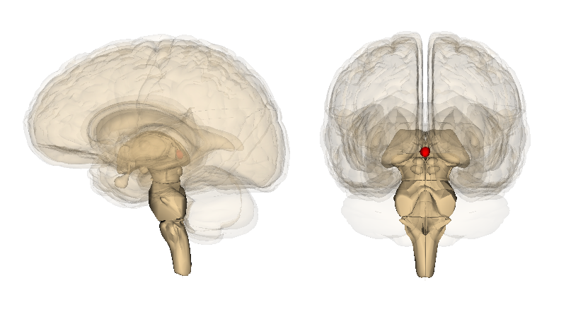
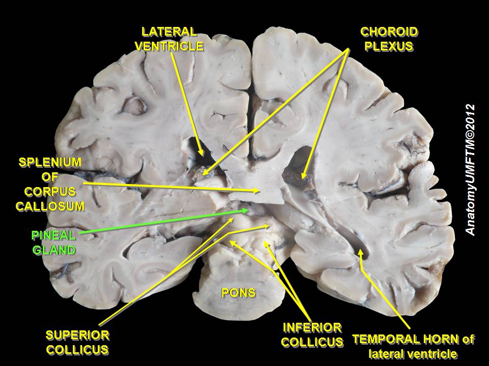
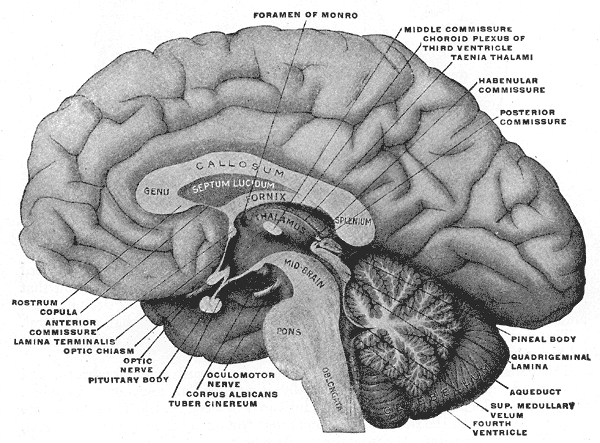
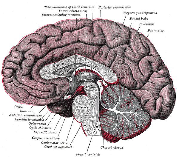
The pineal gland is a small, pine-cone shaped endocrine gland located near the center of the brain, between the two hemispheres where the two halves of the thalamus join. It is present across many vertebrate species and varies in size, position and light-sensitivity between animals; see studies of non-mammalian vertebrates for examples. In humans the gland sits in the epithalamus and is sometimes referred to by older anatomical names such as the epiphysis cerebri or conarium.
Image gallery
10 Images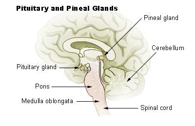



Anatomy and neural connections
Anatomically the pineal gland is compact and richly supplied by blood vessels and sympathetic nerve fibers. It receives information about environmental illumination indirectly from the retina: light perceived by the eyes is transmitted to the brain’s master clock, the suprachiasmatic nucleus, which then influences pineal activity via a multisynaptic pathway. This network of connections allows the gland to translate patterns of external light and darkness into hormonal signals.
Function: melatonin and circadian timing
The gland’s principal hormone is melatonin, released in a daily rhythm with low levels during the day and elevated secretion at night. Melatonin appears in the blood after dusk, promotes the onset of sleepiness in many nocturnal-sleeping species including humans, and falls again toward morning. Through these changes it acts as a biochemical signal of night length and contributes to the timing of sleep, seasonal physiology and some reproductive processes in animals.
Clinical significance and disorders
Because the pineal–melatonin system is central to daily timing, it is implicated in several conditions and therapeutic strategies. Melatonin supplements are used to treat jet lag, shift-work sleep problems and some forms of insomnia. The gland itself can be affected by calcification with age, by benign cysts, or by rare tumours; these lesions can disrupt hormone production or cause pressure effects. Dysregulation of timing mechanisms is linked to a range of problems, including circadian rhythm sleep disorders such as delayed sleep phase, in which the habitual sleep period is consistently pushed to late hours.
Uses, examples and diagnostic notes
- Melatonin therapy: used short-term for jet lag and certain timing disorders; dosing and effectiveness vary by individual.
- Imaging: calcified pineal tissue often shows up on radiographs and CT scanning and can be a useful anatomical landmark.
- Pediatric effects: some pineal tumours influence puberty and endocrine balance and therefore require specialized evaluation.
History and notable facts
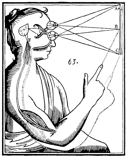
The pineal gland has a notable cultural and philosophical history: it was singled out by some thinkers as a seat of consciousness or soul. Scientific understanding began to coalesce in the 20th century with identification of melatonin and delineation of the light–brain–pineal pathway. In certain reptiles and fish a related structure, the parietal or "third" eye, has direct photoreceptive function, illustrating the evolutionary diversity of this region.
For further reading on anatomy, physiology and clinical aspects see general neuroendocrine summaries and specialty reviews: endocrine gland summaries, imaging and surgical references, and circadian medicine resources available through clinical literature collections. Additional introductory and patient-oriented materials can be found via resources on brain anatomy (middle of the brain) and comparative vertebrate biology (vertebrates).
Related articles
Author
AlegsaOnline.com Pineal gland: structure, function and clinical relevance Leandro Alegsa
URL: https://en.alegsaonline.com/art/76969