
Lacrimal bone — anatomy, function, and clinical relevance
Small, thin facial bone forming part of the medial orbital wall; contributes to tear drainage and the nasolacrimal canal. Important in orbital anatomy, development, and certain eye–nose clinical conditions.
Overview
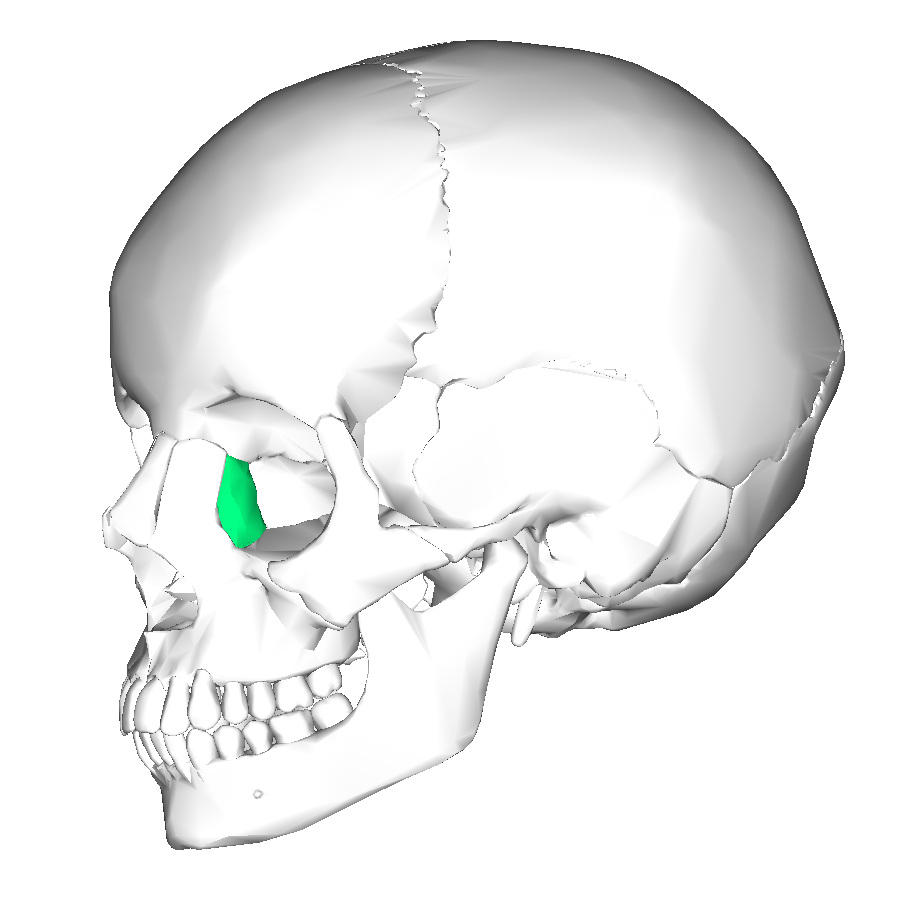
The lacrimal bone is a small, thin bone located in the medial wall of the orbit (eye socket). It is one of the smallest bones of the skull and plays a key structural role in the region where the eye and the nasal cavity meet. Its name comes from the Latin lacrima, meaning "tear," reflecting its relationship to the tear drainage system.
Image gallery
8 Images




Anatomy and features
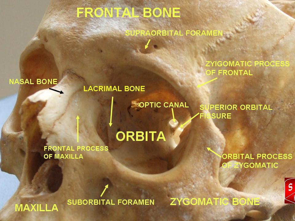
Despite its modest size, the lacrimal bone has several identifiable surfaces and margins and contributes to the formation of the nasolacrimal canal. It typically articulates with adjacent bones such as the frontal bone, maxilla and the ethmoid. The bone is thin and fragile compared with many other cranial bones.
- Lacrimal groove/fossa: a depression that, together with the maxilla, forms the nasolacrimal canal that conducts tears from the lacrimal sac to the nasal cavity.
- Posterior and anterior lacrimal crests: ridges that serve as attachment points for surrounding soft tissues and contribute to the orbital rim.
- Orbital and nasal surfaces: the two faces of the bone that border the eye socket and the nasal cavity respectively.
Development and name
The lacrimal bone develops through intramembranous ossification like many facial bones. Its small size and delicate shape can vary somewhat between individuals. Historically and etymologically, the name reflects its proximity to the tear drainage apparatus rather than any active role in tear production.
Function and clinical relevance
Functionally, the lacrimal bone helps form the bony channel that allows tears to drain from the eye into the nose; damage to this area can interfere with normal tear flow. Clinically, it is relevant in orbital fractures, congenital obstructions of the nasolacrimal duct, and inflammatory conditions of the lacrimal sac such as dacryocystitis.
Examples and notable facts
Surgical procedures addressing blocked tear drainage, for example dacryocystorhinostomy, are performed near the lacrimal bone to create an alternate pathway into the nasal cavity. Because of its small, thin nature it is sometimes fractured in blunt orbital trauma, which can produce persistent tearing (epiphora). For further basic reference see additional anatomical resources.
Related articles
Author
AlegsaOnline.com Lacrimal bone — anatomy, function, and clinical relevance Leandro Alegsa
URL: https://en.alegsaonline.com/art/55279
Sources
- ncbi.nlm.nih.gov : "Diagnostic imaging of the nasolacrimal drainage system. Part I. Radiological anatomy of lacrimal pathways. Physiology of tear secretion and tear outflow"
- doi.org : 10.12659/MSM.890098
- ncbi.nlm.nih.gov : 3999077
- pubmed.ncbi.nlm.nih.gov : 24743297