
Intracranial aneurysm (brain aneurysm): causes, signs, diagnosis, treatment
Intracranial aneurysm: a focal dilation of a cerebral artery. Covers types, causes, clinical presentation, diagnostic imaging, emergency care, treatment options, complications and prevention.
Overview
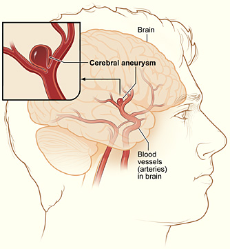
An intracranial aneurysm, commonly called a brain or cerebral aneurysm, is a localized dilation of the wall of an artery inside the skull. The dilation results from a weakness in one or more layers of the arterial wall and can form various shapes and sizes. Many aneurysms remain small and asymptomatic for years; others grow or rupture, releasing blood into the subarachnoid space and causing a form of hemorrhagic stroke that may be life‑threatening or leave lasting disability.
Image gallery
4 Images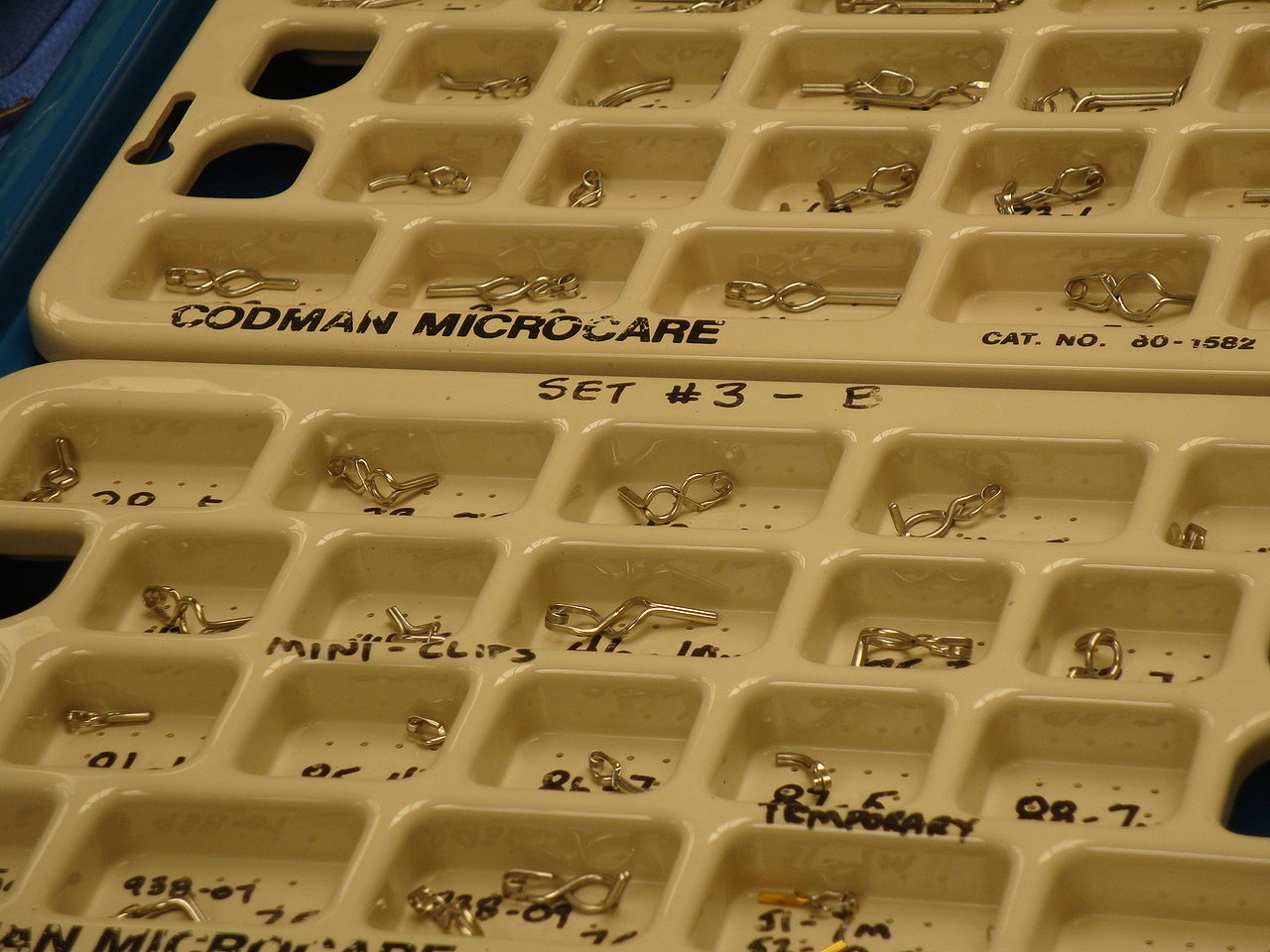


Types and pathophysiology
Aneurysms are described by shape and mechanism. Saccular ("berry") aneurysms are the most commonly discussed type and typically arise at arterial branch points of the circle of Willis and other major intracranial vessels. Fusiform aneurysms involve an elongated, circumferential widening of the vessel. Dissecting aneurysms follow a tear within the arterial wall layers. Infectious (mycotic) aneurysms, traumatic pseudoaneurysms and lesions associated with connective tissue disorders represent other categories. The combination of hemodynamic stress and structural wall weakness contributes to formation and potential growth.
Causes and risk factors
The precise cause of an individual aneurysm often cannot be determined. Contributing factors include chronic high blood pressure, tobacco use, advancing age and inherited conditions that affect connective tissue or vessel integrity, such as autosomal dominant polycystic kidney disease and some collagen disorders. A family history of intracranial aneurysms increases relative risk, and prior head injury or arterial infection can be associated in specific cases. For background on vessel structure and related mechanisms, see blood vessel anatomy and weakness.
Clinical presentation
Unruptured aneurysms are frequently incidental findings on imaging performed for other reasons. When they produce symptoms, this is usually from local compression of adjacent nerves or brain tissue and may cause headaches, double vision, pain above or behind an eye, or focal neurological deficits. Some patients experience a short, intense headache or transient symptoms from a tiny leak (a warning or "sentinel" event) before a major rupture.
- Rupture typically causes sudden, severe headache often described as the worst headache of life, with nausea, vomiting, neck stiffness, and varying levels of reduced consciousness.
- Other acute features can include seizures, focal weakness, speech problems or visual disturbance depending on bleed location.
Diagnosis
Initial assessment of suspected rupture begins with a noncontrast CT scan to detect acute subarachnoid blood. If CT is normal but suspicion remains high, lumbar puncture may identify blood products in cerebrospinal fluid. Vascular imaging defines the aneurysm: CT angiography (CTA) and MR angiography (MRA) are widely used noninvasive tests, while digital subtraction angiography provides the most detailed map for complex cases and treatment planning. For further information on imaging choices, see diagnostic imaging options.
Acute management and complications
A ruptured aneurysm is a neurosurgical emergency. Immediate goals are to stabilize airway, breathing and circulation, manage intracranial pressure, and reduce the risk of rebleeding. Patients are typically treated in an intensive care setting. Common, potentially serious complications after subarachnoid hemorrhage include delayed cerebral ischemia from vasospasm, hydrocephalus requiring cerebrospinal fluid diversion, seizures, and medical complications such as infections or cardiac disturbance. Specific therapies for preventing or treating vasospasm, and supportive interventions for hydrocephalus, are important parts of care.
Definitive treatment options
Definitive exclusion of the aneurysm from the circulation prevents rebleeding. Two principal strategies are used.
- Surgical clipping: an open cranial procedure that places a permanent clip at the aneurysm neck to stop blood flow into the sac.
- Endovascular techniques: catheter-based approaches introduced through blood vessels to deploy coils, stents, or flow‑diverting devices that occlude or remodel the aneurysm from within the artery.
Choice of approach depends on aneurysm anatomy, patient health and available expertise. Some small, low‑risk unruptured aneurysms may be managed conservatively with periodic imaging. For summaries of therapeutic approaches, see treatment approaches.
Prevention, follow‑up and prognosis
Prevention focuses on managing modifiable risks: blood pressure control, smoking cessation, and treatment of other vascular risk factors. When an aneurysm is repaired, follow‑up imaging is commonly recommended to confirm durable occlusion and to monitor for recurrence or the emergence of other lesions. Prognosis varies: many unruptured aneurysms remain clinically silent, but ruptured aneurysms carry substantial risk of death and long‑term disability. Early recognition and rapid treatment improve outcomes.
Screening and special considerations
Screening decisions are individualized. First‑degree relatives of patients with aneurysms, people with certain genetic conditions, and those with a prior history of aneurysmal subarachnoid hemorrhage may be offered selective imaging surveillance. Management during pregnancy, in children, or in patients with multiple medical problems requires multidisciplinary planning.
Resources
Individuals and families often benefit from specialist neurosurgical and neurological consultation, as well as support resources for recovery and rehabilitation. For further patient information and support services, see further information and support.
Related articles
Author
AlegsaOnline.com Intracranial aneurysm (brain aneurysm): causes, signs, diagnosis, treatment Leandro Alegsa
URL: https://en.alegsaonline.com/art/47869