
Coccyx (Tailbone): Structure, Function, and Clinical Significance
Small triangular bone at the base of the spine formed by fused vertebrae; provides pelvic floor and sitting support, site of muscle attachments, and a focus of coccydynia and childbirth-related injury.
The coccyx, commonly called the tailbone, is a small, triangular bony structure located at the lowest end of the vertebral column. It is formed by the fusion of several rudimentary vertebrae and sits below the sacrum. Though much reduced compared with tails in other animals, the coccyx remains an important anatomical landmark and a site for muscular and ligamentous attachments.
Image gallery
3 Images
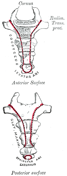

Anatomy and parts
Most human coccyges are composed of three to five tiny vertebral segments that are variably fused. The uppermost part articulates with the sacrum at the sacrococcygeal joint, which allows limited movement in some people. Surface features include a rounded tip pointing downward and pairs of small transverse processes on the fused segments. Several ligaments and muscles attach to the coccyx, helping form the floor of the pelvis.
Attachments and relationships
Important soft-tissue connections include:
- Pelvic floor muscles (such as portions of the levator ani and coccygeus), which support pelvic organs.
- The gluteus maximus, which partly inserts near the coccyx and contributes to hip extension.
- Ligaments, including the anococcygeal ligament and sacrococcygeal ligaments, which stabilize the joint with the sacrum.
Development and evolutionary context
At birth the coccygeal segments are separate ossified pieces that often fuse during adolescence or adulthood. Embryologically the coccyx represents the remnant of a tail—the vertebral column extends beyond the anus in early development—making it a vestigial structure in humans. The degree and timing of fusion vary between individuals.
Function and clinical importance
Although the coccyx no longer serves as a tail, it contributes to weight-bearing when sitting, particularly when leaning back, and provides anchoring points for muscles and ligaments that maintain continence and support pelvic organs. Clinically it is most often discussed in the context of coccydynia (pain in the coccyx), which may result from trauma, repetitive strain, childbirth-related injury, or degenerative change. Diagnosis can involve clinical examination and imaging, and treatment ranges from conservative measures to, rarely, surgical removal (coccygectomy).
Common conditions and notable facts
Conditions associated with the coccyx include bruising, fracture, subluxation of the sacrococcygeal joint, and pain after childbirth. There are known anatomical variations in shape, angle and mobility that influence symptoms and treatment response. For further clinical details and imaging considerations see medical reference.
Distinctions: women often have a more posteriorly angled and more mobile coccyx than men, which can influence childbirth mechanics and susceptibility to pain. Despite being small, the coccyx plays a nontrivial role in pelvic stability and human biomechanics.
Related articles
Author
AlegsaOnline.com Coccyx (Tailbone): Structure, Function, and Clinical Significance Leandro Alegsa
URL: https://en.alegsaonline.com/art/21313