
Blood film (peripheral blood smear)
A blood film is a stained thin smear of peripheral blood on a microscope slide used to evaluate red cells, white cells, platelets and to detect blood parasites and morphological abnormalities.
A blood film, often called a peripheral blood smear, is a thin layer of anticoagulated blood spread across a microscope slide and stained so individual cells can be observed under light microscopy. Properly prepared and stained films permit assessment of red blood cell shape and size, white blood cell differentials, platelet number and appearance, and occasionally the presence of microorganisms or parasites in the bloodstream. A blood film remains a fundamental, low-cost diagnostic tool that supplements automated laboratory analyzers.
Image gallery
8 Images


Preparation and types
Two common formats are the thin film and the thick film. A thin film provides a single layer of cells for detailed morphology and cell differentials and is made by spreading a drop of blood with a second slide. A thick film concentrates blood components by using more blood and allowing some hemolysis; it is more sensitive for detecting small numbers of parasites. The smear is fixed and stained with Romanowsky-type stains to provide contrast between cellular components.
Stains and microscopic features
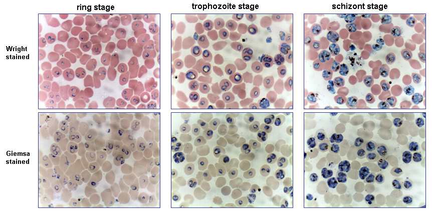
Typical staining methods include Wright, Giemsa and Leishman stains (all Romanowsky variants). Staining highlights nuclei, cytoplasm, granules, and inclusion bodies; it also helps identify developmental stages of parasites. On a stained smear, observers evaluate red cell color and size (anisocytosis, microcytosis, macrocytosis), the presence of poikilocytes (abnormally shaped cells), white blood cell morphology and differential counts, platelet estimates, and any abnormal or immature forms.
Clinical uses and examples
Blood films are used to investigate anemia, infections, hematologic malignancies, and platelet disorders. They can detect parasites in the blood; for example, films are examined specifically when malaria is suspected. Films are also used to confirm or investigate abnormal automated cell counters and to identify diagnostic clues such as blast cells in leukemia or schistocytes in microangiopathic hemolysis. For resources on slide preparation see microscope slide technique.
Advantages, limitations and safety
- Advantages: inexpensive, direct visual assessment, sensitive for certain parasites and cell inclusions.
- Limitations: operator dependent, variable quality of smears and stains, limited quantitative precision compared with automated methods.
- Safety: standard precautions are required to handle potentially infectious blood; proper fixation reduces biohazard risk.
In tropical and travel medicine the film is a frontline test to look for blood parasites and specifically to diagnose malaria. Skilled microscopic interpretation remains essential in settings where automated equipment is unavailable or when unusual cell morphology must be recognized by an experienced observer.
Related articles
Author
AlegsaOnline.com Blood film (peripheral blood smear) Leandro Alegsa
URL: https://en.alegsaonline.com/art/12250