Phalanges: Structure, Function, and Clinical Significance
Phalanges are the small bones of the fingers and toes. This article explains their anatomy, development, roles in hand and foot function, common injuries and variations, and notable distinctions.
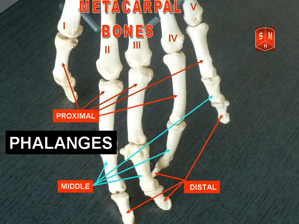
Phalanges are the bones that form the fingers and toes. Each human hand contains 14 phalanges and each foot contains 14 as well. The term describes three general types of segments — proximal, middle (intermediate) and distal — though the thumb and the big toe normally lack a middle phalanx. Phalanges are true bones that articulate with the metacarpals or metatarsals and with one another to enable movement.
Image gallery
10 Images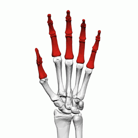


Basic anatomy
In each digit (except the thumb and large toe) there are three phalanges: the proximal phalanx nearest the palm or sole, the middle phalanx, and the distal phalanx at the tip. Joints between them include the proximal and distal interphalangeal joints and the metacarpophalangeal or metatarsophalangeal joints. Tendons for flexion and extension attach to phalanges — for example, flexor tendons insert on the palmar surfaces of the distal and middle phalanges — allowing grasping, manipulation and toe-off during walking. For hand anatomy details see references on the human hand.
Development and evolution
Phalanges form by endochondral ossification from primary and secondary centers during childhood and adolescence; growth plates close as maturity is reached. Vertebrate phalanges are modifications of the ancestral pentadactyl limb and show variation across species in number, proportion and mobility. Human toes are generally shorter and stouter than finger phalanges because of their role in weight-bearing.
Functions and importance
Phalanges contribute to fine motor skills (precision grip, tool use) and to balance and propulsion in locomotion. The arrangement of multiple small bones with joints and tendons gives the digits both strength and flexibility. Distal phalanges support the fingernail and bear sensation-rich tissues that aid touch.
Clinical considerations
Common problems include fractures (tuft, shaft, base), joint disorders (osteoarthritis, inflammatory arthritis), deformities (mallet finger, hammer toe, clinodactyly) and congenital variations (polydactyly or fused phalanges). Treatment ranges from splinting to surgical fixation and joint reconstruction. Foot-specific conditions often affect the toes and gait; for foot and toe issues consult resources about the foot.
- Key joints: metacarpophalangeal, proximal interphalangeal, distal interphalangeal.
- Typical variation: thumb and great toe each have two phalanges instead of three.
- Clinical imaging: x-rays are the primary method to assess phalangeal fractures and alignment.
Understanding phalangeal structure helps explain everyday functions like gripping and walking, as well as many common injuries and congenital differences. For more detailed anatomical schematics and clinical guidelines, consult specialized anatomy and orthopedics sources or the linked references above.
Related articles
Author
AlegsaOnline.com Phalanges: Structure, Function, and Clinical Significance Leandro Alegsa
URL: https://en.alegsaonline.com/art/76324