Microanatomy: the microscopic structure of tissues and cells
Microanatomy examines the structures of cells, tissues and organ components using microscopes and preparation techniques; it underpins pathology, developmental biology and clinical diagnosis.
Microanatomy refers to the study of biological structures that are too small to be resolved with the naked eye. It is a subordinate discipline of anatomy that focuses on the organization and relationships of cells, extracellular matrix, and the specialized arrangements within organs. When the emphasis is on tissues and their cellular constituents, the term histology is commonly used; cytology addresses individual cell form and content.
Image gallery
10 Images


Methods and tools
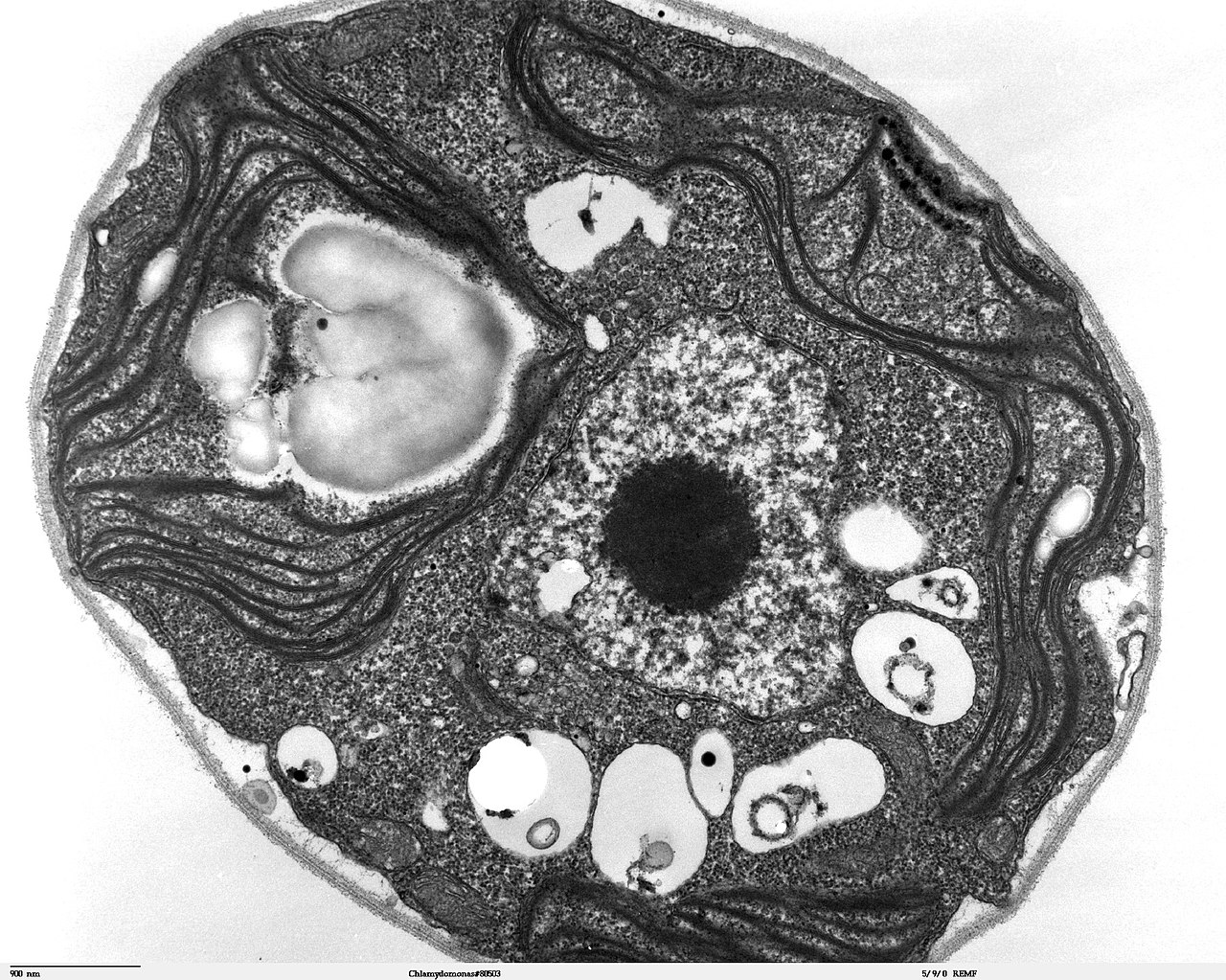
Investigators in microanatomy use a range of instruments and preparation methods to reveal structure. Routine approaches include light microscopy of thin sections, special staining to highlight particular components, and transmission or scanning electron microscopy for ultrastructural detail. Tissue processing—fixation, embedding, sectioning and staining—creates the thin slices required for observation, while more recent techniques such as immunohistochemistry allow selective visualization of molecules within cells.
Typical scales of interest run from organelles within cells (nanometres to micrometres) up to the architecture of tissue layers within an organ (tens to hundreds of micrometres). Common tissue classes examined are epithelial, connective, muscle and nervous tissues, each with characteristic cell types and arrangements that relate to function.
Applications and importance
Microanatomy is fundamental to biomedical science and clinical practice. It underlies diagnostic pathology—examining biopsies to detect disease, infection or neoplasia—and supports research in development, physiology and toxicology. Examples of routine applications include identifying inflammatory patterns, classifying tumor types, and tracing the effects of drugs on cellular structure. Because structure and function are tightly linked at microscopic scales, observations in microanatomy frequently inform physiological and clinical interpretation.
Key distinctions and considerations include:
- Levels of focus: cytology (single cells), histology (tissues), and ultrastructure (organelles).
- Resolution trade-offs: light microscopy offers color and context over larger areas; electron microscopy provides far greater detail but requires more complex preparation.
- Limitations: artefacts from processing and subjective interpretation can affect conclusions, so multiple methods are often combined.
Historically, microanatomy advanced with improvements in lenses and staining in the 17th–19th centuries and later with the development of electron microscopy and molecular labeling methods in the 20th century. Today it remains a dynamic field, integrating classical observation with molecular and digital imaging tools to deepen understanding of normal structure and disease-related change.
Related articles
Author
AlegsaOnline.com Microanatomy: the microscopic structure of tissues and cells Leandro Alegsa
URL: https://en.alegsaonline.com/art/64585
Sources
- medical-dictionary.thefreedictionary.com : "Microanatomy"
- dictionary.com : "The Definition of Microanatomy"