Lymph node
Small, encapsulated immune organs that filter lymph and coordinate immune responses; located throughout the body and important in infection control, cancer staging, and immune surveillance.
Lymph nodes are small, bean-shaped organs of the lymphatic system that act as filters and coordination centers for immune responses. Encapsulated and highly organized, each node houses a variety of immune cells that monitor lymph fluid for pathogens, foreign particles and abnormal cells. The nodes are essential for adaptive immunity: they support interactions between antigen-presenting cells, B lymphocytes and T lymphocytes to produce targeted responses and immunological memory. For a simple reference to the range of cell types typically present in nodes, see basic immunology resources.
Image gallery
9 Images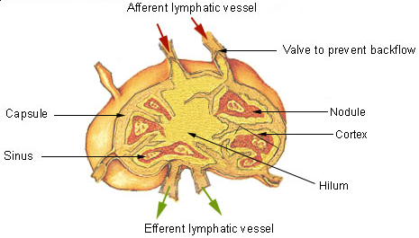


Structure and components
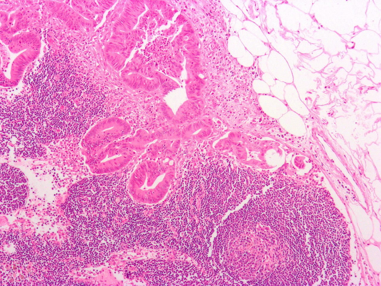
Internally a lymph node is divided into regions with different roles. The outer cortex contains follicles rich in B cells and, when activated, germinal centers where antibody-producing cells mature. The paracortex is T-cell–rich and is the main site for antigen presentation and T-cell activation. The medulla contains plasma cells and macrophages and leads to efferent lymphatic vessels that carry filtered lymph away. A surrounding fibrous capsule and a system of trabeculae support the organ while sinuses allow lymph to percolate past immune cells. Many introductory texts describe these compartments and their cellular inhabitants; for context on circulating white blood cells and their behavior, consult immunology guides.
Function and roles
Lymph nodes perform several complementary functions:
- Filtering lymph: trapping microbes, debris and foreign particles carried from tissues.
- Antigen presentation: dendritic cells bring antigens into the node and present them to lymphocytes.
- Immune activation and amplification: B and T cells proliferate and differentiate in response to specific antigens.
- Generating memory: formation of memory B and T cells that confer long-term protection.
These processes make nodes central to the body's immune function, enabling localized detection and systemic responses to infection and injury.
Distribution and clinical importance
Lymph nodes are grouped in predictable regions such as cervical (neck), axillary (armpit), inguinal (groin), and mesenteric (abdominal) chains. Their pattern of drainage is clinically useful: swollen (tender) nodes commonly indicate local infection, while hard, fixed nodes may raise concern for malignancy. Physicians use the presence, size and consistency of nodes in diagnosis, and surgeons perform sentinel node biopsies to stage certain cancers. Persistent generalized enlargement is termed lymphadenopathy and can have infectious, inflammatory, or neoplastic causes.
Origin, development and distinctions
Lymph nodes develop during fetal life as components of the growing lymphatic network, arising from interactions between lymphatic endothelial cells and adjacent stromal cells. Historically, anatomists and immunologists gradually elucidated node structure and function as microscopy and cellular biology advanced. It is important to distinguish lymph nodes from other lymphoid organs: the spleen filters blood rather than lymph, while tonsils and Peyer patches sample antigens at mucosal surfaces without the same encapsulated architecture. For an overview of the broader lymphatic system, consult standard anatomy references.
Because of their central role in immune surveillance, lymph nodes remain a major focus in medicine and research—from understanding infection dynamics to improving cancer staging and designing vaccines that induce robust lymph-node–based immune responses.
Related articles
Author
AlegsaOnline.com Lymph node Leandro Alegsa
URL: https://en.alegsaonline.com/art/60101
Sources
- thelymphnodes.com : "Lymph Node"