Interstitial fluid
Clear extracellular fluid that fills tissue spaces between cells, transporting nutrients, gases and wastes; essential for cell environment, fluid balance, immunity and clinical conditions.
Overview
Interstitial fluid is the clear, lightly proteinaceous fluid that occupies the small spaces between cells in tissues. It is the major component of the extracellular fluid compartment and — in many descriptions — comprises about three quarters of extracellular fluid. Interstitial fluid acts as the immediate environment through which cells receive nutrients, exchange gases and eliminate metabolic wastes. For a general introduction see interstitial fluid overview.
Image gallery
3 Images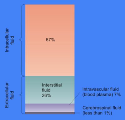


Composition and distribution
The chemical composition of interstitial fluid broadly resembles that of blood plasma but differs in concentration of large proteins: it has lower levels of plasma proteins and a different mix of signaling molecules. Major dissolved constituents include electrolytes (sodium, chloride, bicarbonate), small organic nutrients (glucose, amino acids), dissolved gases and locally produced metabolites. Immune cells and signaling peptides travel through this space, enabling inflammation and immune surveillance. Specialized interstitial-like fluids in particular sites include synovial fluid in joints, intraocular fluid in the eye, and serous fluids such as pericardial or pleural fluid; see related compartments: plasmatic, lymphatic and synovial.
Formation and clearance
Interstitial fluid is produced mainly by filtration across capillary walls. Hydrostatic pressure in capillaries pushes water and small solutes into the interstitium, while plasma oncotic pressure (largely due to albumin) opposes that movement. Excess interstitial fluid is returned to the circulation via the lymphatic system, which collects fluid and macromolecules and eventually drains into central veins. The balance of formation and clearance maintains tissue hydration and steady solute concentrations; basic mechanisms are discussed in summaries of tissue fluid exchange: exchange mechanisms.
Functions
- Transport of nutrients, gases and metabolic wastes between blood and cells.
- Facilitation of intercellular signaling by distributing hormones, cytokines and growth factors.
- Support for immune surveillance by allowing leukocyte trafficking and antigen transport to lymphatics.
- Mechanical buffering of cells and contribution to local ionic and pH homeostasis.
Clinical significance
Disruption of the normal balance between production and removal of interstitial fluid leads to clinical problems. Excess accumulation produces edema, while impaired generation or altered composition can affect tissue function. Common contributors to edema include heart failure, kidney disease, low plasma protein states (hypoalbuminemia), inflammation and lymphatic obstruction. Abnormal effusions of serous cavities (pleural, pericardial, peritoneal) are related phenomena and are assessed in clinical practice by physical exam, imaging and fluid analysis; see summaries of fluid compartments and clinical implications: extracellular fluid fractions.
Distinctions and examples
- Interstitial fluid versus plasma: lower protein concentration and different colloid osmotic pressure.
- Interstitial versus intracellular fluid: distinct ionic makeup and tightly regulated cell volume mechanisms.
- Local adaptations: synovial fluid is specialized for lubrication, intraocular fluids support eye shape and optics, and serous fluids reduce friction around organs.
For concise primers and more detailed reviews consult general overviews and specialty material on microcirculation, lymphatics and joint physiology: general overviews, exchange mechanisms, fluid compartments, plasma relations, lymphatics and joint fluid.
Author
AlegsaOnline.com Interstitial fluid Leandro Alegsa
URL: https://en.alegsaonline.com/art/47841