
Electroencephalography (EEG): recording and interpreting brain electrical activity
Electroencephalography (EEG) is a noninvasive method that records electrical activity of the brain from the scalp. It aids diagnosis of seizures, sleep disorders, coma and brain function, and supports research into brain rhythms.
Overview
Electroencephalography (EEG) records electrical patterns produced by the brain at the scalp. It provides a continuous, noninvasive measure of brain activity by detecting tiny voltage fluctuations generated when populations of nerve cells communicate using ionic currents and electricity. Those coordinated signals carry information about sensory processing, movement, sleep and cognition. An EEG recording, called an electroencephalogram, is a primary tool in neurological diagnosis and research.
Image gallery
9 Images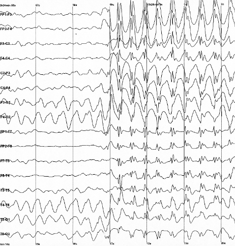



How EEG works and equipment
Surface electrodes are placed on the scalp according to standardized layouts (for example the 10–20 system) and connected to differential amplifiers and a recorder. Modern systems digitize the signal at high sampling rates and apply filters to reduce noise. Preparation includes cleaning the skin and applying conductive paste or gel; recordings can be short (routine 20–30 minutes) or long (ambulatory or continuous monitoring for days). Montage selection, electrode placement and filter settings affect the appearance of the traces and the ability to localize sources. Specialized setups add synchronized video, event markers or stimulus delivery for evoked potentials.
Characteristics and common rhythms
EEG waveforms are described by frequency, amplitude and distribution across the head. Common bands are delta (0.5–4 Hz; large slow waves seen in deep sleep or pathology), theta (4–8 Hz), alpha (8–13 Hz; often strongest over posterior regions and enhanced with eyes closed), beta (13–30 Hz; associated with alert activity), and gamma (above 30 Hz; related to high-frequency processes). Clinicians must distinguish physiological rhythms from artifacts produced by muscles, eye movements, cardiac activity, poor electrode contact or external electrical devices. Quantitative measures and topographic maps are sometimes used to summarize patterns across many channels.
History and development
Recordings of human scalp electrical activity date to the early 20th century, with systematic human studies performed by physicians such as Hans Berger. Progress in amplifier design, electrode materials, video synchronization and digital signal processing over the 20th and 21st centuries expanded EEG from a niche research tool into a routine clinical modality. Contemporary advances include high-density electrode caps, computerized quantitative analyses and integration with other modalities to improve spatial localization and diagnostic utility.
Uses, examples and importance
EEG is essential in many clinical settings. Typical uses include:
- Evaluation and classification of seizures and epilepsy, including localization of seizure onset for surgical planning.
- Continuous monitoring in intensive care units to assess encephalopathy, detect nonconvulsive seizures and inform prognosis in coma or after brain injury.
- Sleep studies that use EEG to stage sleep, measure sleep rhythms and diagnose disorders such as sleep apnea and narcolepsy.
- Confirmation of brain death under specific protocols, assessment of neonatal brain function, and monitoring during epilepsy surgery.
EEG also supports research into cognition, brain–computer interfaces and neurodevelopment. Techniques such as evoked potentials extract responses time-locked to stimuli, improving sensitivity for specific pathways. Ambulatory and video-EEG extend diagnostic yield by capturing rare events in natural settings.
Limitations, variants and practical considerations
Although EEG offers excellent temporal resolution, its ability to pinpoint deep or small cortical sources is limited; spatial resolution is improved with intracranial recordings or by combining EEG with MRI. Interpretation requires trained specialists because many findings are nonspecific. Artifacts, patient movement and technical faults can obscure results. Variants include high-density EEG, ambulatory long-term recording, intracranial EEG for presurgical evaluation, quantitative EEG analyses and simultaneous multimodal recordings (for example EEG-fMRI). When used appropriately, EEG provides unique real-time insight into brain function but usually complements other clinical and imaging data.
Author
AlegsaOnline.com Electroencephalography (EEG): recording and interpreting brain electrical activity Leandro Alegsa
URL: https://en.alegsaonline.com/art/30718