
Cerebral infarction
Cerebral infarction is brain tissue necrosis caused by prolonged loss of blood flow (ischemia). This article explains causes, types, clinical features, diagnosis, treatment, prognosis, and prevention.
Overview
A cerebral infarction is an area of dead (necrotic) brain tissue resulting from an interruption of the blood supply. When blood flow and oxygen delivery fall below the level needed to maintain cellular metabolism, neurons and supporting cells suffer irreversible injury. This process is a form of ischemic stroke and may follow a brief period of impaired perfusion or a complete and sustained blockage. In medical descriptions, damaged tissue is often referred to as infarcted brain tissue.
Image gallery
4 Images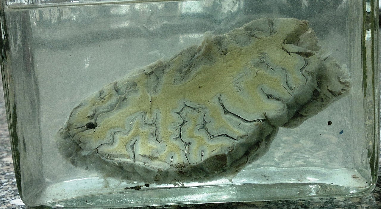

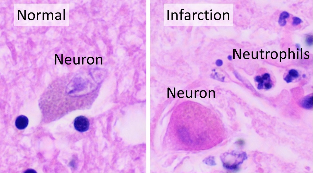

Causes and mechanism
Most cerebral infarctions arise when an arterial supply is narrowed or occluded. Common mechanisms include local clot formation (thrombosis), material traveling from elsewhere in the circulation (embolism), and progressive narrowing from atherosclerotic disease. The shortage of oxygen and glucose triggers a cascade of events — loss of ion homeostasis, excitotoxicity, inflammation, and cell death — that enlarges the injured area if flow is not promptly restored. The clinical syndrome produced by this process is described as an ischemic stroke.
Types and regions affected
Infarcts are often classified by size and arterial territory. Territorial infarcts involve the distribution of a main cerebral artery, such as the middle, anterior, or posterior cerebral arteries. Lacunar infarcts are small, deep lesions affecting subcortical structures and are commonly related to small-vessel disease. Watershed infarcts occur in border zones between arterial territories during systemic low flow. Which artery is involved largely determines the neurologic deficits seen.
Typical symptoms and signs
- Sudden weakness or numbness, often on one side of the body.
- Difficulty speaking or understanding language when language areas are affected.
- Visual changes including field loss if posterior circulation is involved.
- Coordination problems, dizziness, or decreased consciousness in larger or brainstem infarcts.
Diagnosis and acute management
Rapid neuroimaging (noncontrast CT or diffusion-weighted MRI) is used to distinguish ischemia from hemorrhage and to localize the infarct. Acute treatments aim to restore perfusion and limit damage. When appropriate and within time windows defined by clinical guidelines, intravenous thrombolytic medication and endovascular thrombectomy can reopen blocked arteries. Other measures include antiplatelet or anticoagulant therapy for selected causes, blood pressure and glucose management, and supportive care to prevent complications.
Prognosis, prevention, and notable facts
Outcomes vary with infarct size, location, patient age, comorbidity, and speed of treatment. A substantial proportion of ischemic strokes have severe outcomes; historically, around one third have resulted in death, though modern acute care has improved survival and functional recovery for many patients. Rehabilitation is a cornerstone of recovery to regain function. Preventive strategies focus on controlling risk factors such as hypertension, atrial fibrillation, diabetes, smoking, and atherosclerosis of major arteries. When clots form in large arteries or the heart they may travel to the brain; these embolic sources may include cardiac thrombus or thrombosis at a ruptured plaque (thrombus), and identification of the source guides long-term therapy.
For clinicians and patients seeking more detailed protocols and evidence reviews, refer to contemporary stroke guidelines and specialist resources: clinical overview, acute stroke care, and stroke prevention summaries (antithrombotic therapy, vascular risk management).
Author
AlegsaOnline.com Cerebral infarction Leandro Alegsa
URL: https://en.alegsaonline.com/art/18173