
Thrombus (blood clot): formation, structure, and clinical significance
A thrombus is a blood clot formed during coagulation that seals damaged vessels; it can be protective or harmful when it blocks circulation. Overview, composition, clinical roles, and distinctions.
A thrombus, commonly called a blood clot, is an aggregate of blood constituents that forms at a site of vascular injury to limit blood loss. In normal physiology the process helps stop bleeding, but under other circumstances a clot can persist inside a vessel and impair flow. Thrombi vary in size, texture and stability depending on where and how they form.
Image gallery
7 Images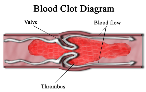



Composition and how thrombi form
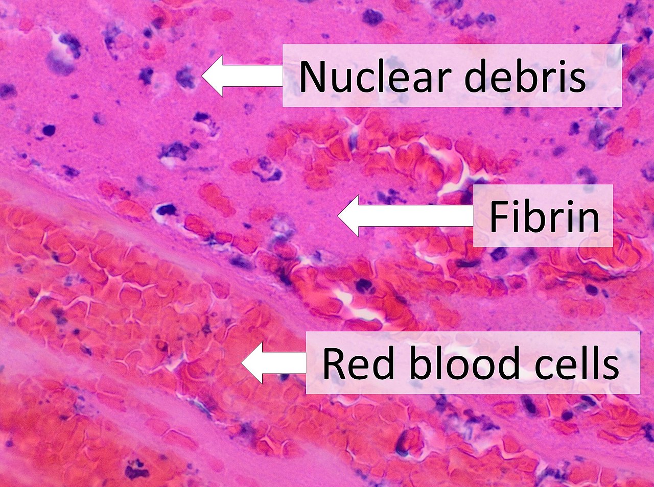
Thrombus formation is the end result of the coagulation cascade and platelets working together to produce a stable plug. Typical components include:
- Aggregated platelets that adhere to the damaged vessel wall.
- Fibrin strands that create a mesh, produced by the coagulation system often referred to as coagulation.
- Entrapped blood cells such as red blood cells and leukocytes.
The material of a thrombus is sometimes called cruor in older texts. Formation typically begins when the endothelial surface is disrupted or when blood flow and composition change in ways that favor clotting.
Physiologic role and pathological consequences
Thrombi serve an essential hemostatic role after injury by sealing breaks in vessels and allowing tissue repair. However, when clots occur inappropriately within intact vessels it is referred to as thrombosis. A pathological thrombus can reduce or block blood flow, causing ischemia of downstream tissues. Pieces may detach and travel as emboli, lodging in distant vessels.
Clinically, thrombi are important in disorders such as deep vein thrombosis, pulmonary embolism, myocardial infarction and ischemic stroke. Diagnosis may use imaging and blood tests of coagulation, while prevention and treatment include anticoagulant and antiplatelet drugs, mechanical measures, or procedures to remove or dissolve clots.
Notable distinctions include the difference between a mural thrombus (attached to a vessel wall), a red thrombus rich in erythrocytes, and a white thrombus rich in platelets. Over time some thrombi can be organized by ingrowth of tissue, become recanalized, or undergo lysis. Understanding when a thrombus is protective and when it becomes pathological is central to preventing and managing cardiovascular and thromboembolic disease.


Types thrombi
By composition
- White thrombus (deposition thrombus): fibrin-rich thrombus
- Red thrombus (clotting thrombus): blood-rich thrombus
- Platelet thrombus: Thrombus consisting predominantly of platelets.
- Fibrin thrombus: Thrombus consisting only of fibrin.
- Phlebolith: also vein stone, a calcified vein thrombus in calcinosis dystrophica
According to place of origin
- Arterial thrombus: mostly wall thrombus formed predominantly by platelets and fibrin.
- Venous thrombus: red thrombus often formed in the area of the valves due to stasis
- Atrial thrombus: atrial fibrillation can lead to thrombus formation in the atrium (especially in the heart ear).

Inhibitors of thrombus formation
- Antiplatelet drugs such as acetylsalicylic acid (ASA; only for arterial thrombi) or clopidogrel
- Heparin
- 4-hydroxycoumarins, e.g. phenprocoumon
- Thrombin antagonists
- Direct oral anticoagulants (DOAKs or DOAKs)
Related articles
Author
AlegsaOnline.com Thrombus (blood clot): formation, structure, and clinical significance Leandro Alegsa
URL: https://en.alegsaonline.com/art/99676
Sources
- worldcat.org : 879416939
- pathophys.org : "Venous thromboembolism (VTE) | McMaster Pathophysiology Review"