
Cerebral arteriovenous malformation (brain AVM)
A cerebral arteriovenous malformation is an abnormal, usually congenital, tangle of arteries and veins in the brain that can cause bleeding, seizures, headaches and focal neurological problems; care is individualized.
Overview
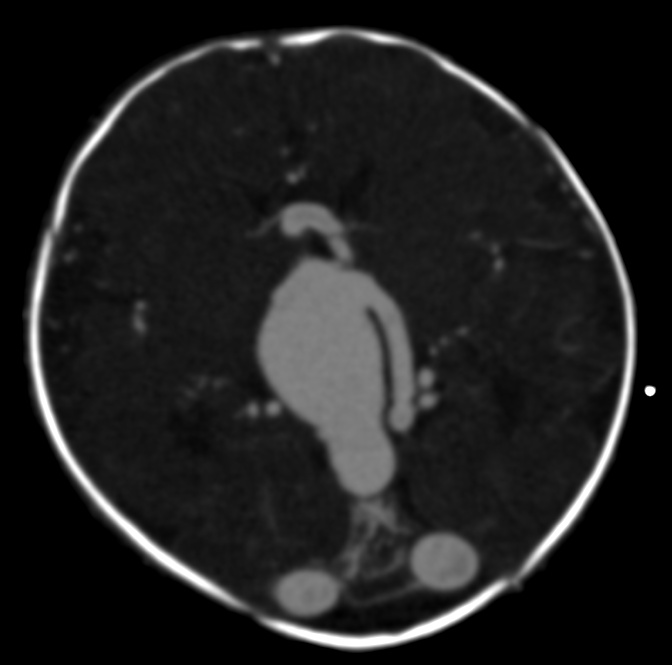
A cerebral arteriovenous malformation (AVM) is an abnormal connection between arteries and veins in the brain that bypasses the normal capillary network. Located in the cerebrum or other intracranial structures, an AVM forms a tangled cluster of vessels called a nidus. It is typically regarded as a developmental vascular lesion that may remain asymptomatic for years or become apparent because of bleeding, seizures or other neurological symptoms.
Image gallery
3 Images

Pathophysiology
In an AVM blood flows directly from high-pressure arteries into low-resistance veins. The absence of capillaries alters local pressure and flow dynamics, which can dilate veins, produce chronic ischemia in nearby brain tissue, and weaken vessel walls. These changes increase the risk of rupture and intracranial hemorrhage, particularly when certain anatomic features are present.
Clinical presentation
Presentation varies by size and location. Common manifestations include:
- Bleeding: sudden headache, vomiting, reduced consciousness or focal deficit due to intracranial bleeding.
- Seizures: new-onset seizures are a frequent initial symptom and may be focal or generalized (seizures).
- Headache and focal deficits: chronic or recurrent headaches, progressive weakness, numbness, speech or visual disturbances depending on the region involved.
- Less common effects: cognitive or behavioral changes, hydrocephalus if bleeding or mass effect obstructs cerebrospinal fluid pathways.
Diagnosis
Imaging is central to diagnosis. CT or MRI can detect hemorrhage, hemosiderin, or an abnormal vascular mass. Digital subtraction angiography (DSA) is the reference standard for detailed mapping of feeding arteries, the nidus and venous drainage and is usually required for treatment planning. Noninvasive vascular imaging helps during follow-up.
Grading and risk assessment
Risk assessment for natural history and treatment commonly uses established grading systems that consider lesion size, location in eloquent cortex, and venous drainage patterns. Prior hemorrhage, deep venous drainage and certain angioarchitectural features are associated with higher risk of future bleeding. Decisions about intervention weigh these factors against procedural risks.
Treatment options
Management is individualized and often decided by multidisciplinary teams. Options include:
- Conservative observation for small, asymptomatic lesions or when treatment risks outweigh benefits.
- Surgical resection to remove the nidus when it is accessible and morbidity is acceptable.
- Endovascular embolization to reduce flow or obliterate portions of the nidus as a standalone therapy or as an adjunct to surgery or radiosurgery.
- Stereotactic radiosurgery for small to moderate lesions in deep or eloquent areas; clinical effect develops over months to years.
Combinations of these modalities are common. The choice depends on anatomy, symptoms and patient preferences.
Complications, prognosis and follow-up
Potential complications include recurrent hemorrhage, persistent seizures, focal neurological deficits, and cognitive effects. Prognosis varies: some AVMs remain stable for years while others carry significant hemorrhagic risk. After treatment, follow-up imaging is required to document obliteration or detect recurrence. Long-term care often involves neurologists, neurosurgeons and rehabilitation specialists.
Special considerations
Pregnancy, anticoagulation and coexisting vascular conditions may influence presentation and management; pregnant patients and those with complex lesions benefit from specialist input. Patients and families are usually advised to seek care at centers with experience in cerebrovascular disorders and multidisciplinary review of imaging and treatment options, including resources on arteriovenous malformation anatomy and clinical management, vascular imaging such as arterial and venous studies, and guidance on hemorrhage and symptom control (intracranial hemorrhage, headache, seizure care).
Author
AlegsaOnline.com Cerebral arteriovenous malformation (brain AVM) Leandro Alegsa
URL: https://en.alegsaonline.com/art/18170
Sources
- doi.org : 10.3171/jns.1966.25.4.0467
- worldcat.org : 0022-3085
- pubmed.ncbi.nlm.nih.gov : 5925721
- surgicalneurologyint.com : "Oculomotor neuropathy from an unruptured arteriovenous malformation in the frontal operculum: A case report"