
Tympanic membrane (eardrum): structure, function and clinical significance
The tympanic membrane, or eardrum, is a thin, semi-transparent membrane separating the external ear from the middle ear that transmits sound vibrations to the ossicles and plays a key role in hearing and ear health.
The tympanic membrane, commonly called the eardrum, is a delicate, conical membrane that forms the boundary between the external auditory canal and the middle ear. It serves as the first mechanical interface for airborne sound, converting pressure waves in air into vibrations that are then relayed inward. For a concise schematic description see structural overview.
Image gallery
10 Images


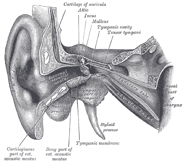
Structure and components
Microscopically the membrane consists of three layers: an outer epidermal layer continuous with the ear canal skin, a fibrous middle layer that provides tensile strength, and an inner mucosal layer facing the middle ear. The central portion, called the pars tensa, is tighter and most important for sound conduction; a smaller, more flaccid region near the top is the pars flaccida. The membrane’s shape and tension affect how it vibrates in response to different frequencies.
Function and mechanical role
When sound reaches the ear canal, the tympanic membrane vibrates and transmits mechanical energy to the middle ear bones. The malleus (hammer) is attached to the inner surface of the membrane and bridges to the incus and stapes; together these three ossicles amplify and convey motion to the inner ear. More on sound transfer and middle ear mechanics is available at sound transmission, and a focused discussion of the ossicular chain is at ossicles and linkage.
Beyond simple transmission, the eardrum contributes to impedance matching between air and the fluid-filled cochlea, enhancing efficiency of hearing across a range of frequencies. Its mobility can be altered by pressure changes, scarring, or fluid in the middle ear, all of which affect hearing acuity.
Clinical importance and common conditions
- Otitis media: middle ear infection often causes fluid accumulation and reduced eardrum mobility.
- Perforation: tears or holes in the membrane from infection, trauma, or barotrauma can reduce hearing and permit recurrent infections.
- Myringosclerosis and scarring: repeated disease or surgery may stiffen the membrane and change its vibratory properties.
Examination with an otoscope provides direct view of the tympanic membrane and remains a primary diagnostic step. Objective tests such as tympanometry measure membrane compliance, while audiometry assesses functional impact. Surgical procedures range from simple patching of small perforations to tympanoplasty to reconstruct larger defects; in some cases ventilation tubes are inserted to manage chronic middle ear effusion.
Across vertebrates, an analogous membrane or tympanum is present in many terrestrial groups and plays comparable roles in hearing, though exact morphology varies by species. Understanding the tympanic membrane’s anatomy and mechanics is fundamental to diagnosing ear disease, planning treatment, and interpreting how sound is transferred from the environment into the inner ear.
Related articles
Author
AlegsaOnline.com Tympanic membrane (eardrum): structure, function and clinical significance Leandro Alegsa
URL: https://en.alegsaonline.com/art/102318
Sources
- britannica.com : "tympanic membrane (anatomy) -- Britannica Online Encyclopedia"