Notochord: embryonic axial structure and evolutionary role
The notochord is a flexible, rod-like axial structure present in all chordate embryos. It provides mechanical support, directs nearby tissue patterning through signaling, and is variably retained or transformed in adults.
Overview
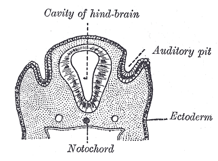
The notochord is a flexible, rod-shaped midline structure present in the embryos of all animals in the phylum Chordata. It lies ventral to the developing neural tube and helps establish the primary body axis during early development. In many vertebrates it is transient, acting as a scaffold while the vertebral column or other skeletal elements form; in some basal chordates it persists into adulthood as the main axial support. For basic context see embryonic development and for taxonomic background see chordates. The developing neural tube and future spinal cord are anatomically and functionally associated with the notochord (spinal cord).
Image gallery
9 Images


Structure and composition
Microscopically, the notochord is composed of large, vacuolated cells surrounded by a sheath of connective tissue and extracellular matrix. The vacuolated cells resist compression while the sheath provides tensile strength, so the combined structure behaves as a hydrostatic or elastic rod: stiff enough to transmit muscular forces, yet flexible enough to permit lateral bending during locomotion. In many descriptions the notochord is therefore called a hydrostatic skeleton or axial rod.
Development and molecular role
The notochord arises early from axial mesodermal tissues and plays a key inductive role in patterning adjacent tissues. It secretes signaling molecules that influence the differentiation of the overlying neural tube and surrounding mesoderm. One well-studied example is the production of morphogens that help specify dorsal–ventral identities within the neural tube and direct the formation of structures such as motor neurons and floor plate cells. As a signalling center it coordinates regional development along the embryo's midline.
Fate in vertebrates and clinical relevance
During vertebrate embryogenesis the vertebral column forms around the notochord; in many species the notochord is largely replaced by vertebral bodies but remnants remain. In mammals notochordal cells contribute to the nucleus pulposus, the gelatinous core of intervertebral discs. Degeneration or loss of function in disc tissues can contribute to back pain and disc herniation, so the notochord's developmental origin is relevant to understanding certain spinal conditions.
Variation across chordates
Comparative anatomy shows a range of outcomes across chordates. In cephalochordates (lancelets) the notochord persists as a primary support throughout life and extends into the head region. In tunicates the notochord is a conspicuous feature of the free-swimming larva and is often reduced or lost in the sessile adult. In most fishes, amphibians, reptiles, birds and mammals the notochord is integrated into the developing axial skeleton and becomes less prominent in the adult.
Functions summarized
- Mechanical support: provides a central rod for muscle attachment and transmits forces for swimming or bending.
- Developmental signaling: secretes diffusible factors that pattern the neural tube and adjacent mesoderm.
- Scaffold for skeleton formation: organizes the vertebral column in vertebrates and contributes cells to intervertebral structures.
Evolutionary significance and research
The notochord is one of the defining characters of chordates and represents an ancestral solution to axial support that predates mineralized vertebrae. Its combination of structural and inductive roles illustrates how a single embryonic feature can be repurposed across evolutionary time. Because of its central role in early patterning and its varying persistence among taxa, the notochord is studied across many model organisms to understand both basic developmental mechanisms and the evolutionary diversification of the chordate body plan. For further reading on developmental and comparative aspects see resources on embryonic development, the diversity of chordates, and the anatomy of the spinal cord.
Related articles
Author
AlegsaOnline.com Notochord: embryonic axial structure and evolutionary role Leandro Alegsa
URL: https://en.alegsaonline.com/art/71172