
Functional magnetic resonance imaging (fMRI): principles, uses, and limitations
fMRI is a noninvasive neuroimaging method that maps brain activity by detecting changes in blood oxygenation (BOLD). It is widely used in research and clinical functional mapping.
Overview
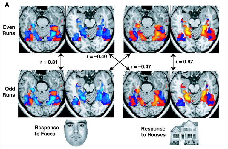
Functional magnetic resonance imaging (fMRI) is a noninvasive technique that produces images reflecting changes in brain activity over time. Rather than visualizing structure alone, fMRI highlights regions where metabolic and vascular changes accompany neural processing. Researchers and clinicians use it to study perception, cognition, language, emotion and to localize critical functions before brain surgery. For a general introduction to how fMRI relates to measured brain processes see brain activity resources.
Image gallery
6 Images




Basic mechanism
The most common fMRI signal is the blood-oxygen-level-dependent (BOLD) contrast. When neurons become active they consume oxygen; the brain responds by increasing local blood flow more than is strictly needed, altering the relative amounts of oxygenated (oxyhemoglobin) and deoxygenated (deoxyhemoglobin) blood. Deoxyhemoglobin has magnetic properties that affect the MRI signal, so local changes in oxygenation produce measurable differences in image intensity. This is why active regions can appear to "light up" on fMRI sequences. Simple explanations of oxygen use and contrast are available at oxygen and imaging and technical discussions about hemoglobin effects can be found at hemoglobin contrast.
Historical notes and development
fMRI grew out of advances in MRI physics and the recognition that blood oxygenation affects magnetic resonance signals. In the early 1990s investigators applied these effects to map functional responses in humans. Since then, both hardware and analysis methods have evolved, improving spatial and temporal resolution and enabling new applications such as resting-state connectivity and multi-voxel pattern analyses. For methodological resources see technical fMRI guides.
Common applications
- Basic cognitive and systems neuroscience research (memory, attention, perception).
- Clinical mapping of language, motor and sensory areas before neurosurgery.
- Studies of functional connectivity and network organization in health and disease.
- Pharmacological and developmental studies that track changes over time.
Strengths and limitations
fMRI offers good spatial detail (millimeter scale) and broad brain coverage without ionizing radiation, making it suitable for repeated measures. However, it measures vascular correlates of neural activity, not electrical firing directly, so interpretations must account for the hemodynamic delay and indirect nature of the signal. Typical hemodynamic responses evolve over seconds, limiting temporal precision. The technique is also sensitive to subject motion, physiological noise, and magnetic susceptibility artifacts, and it requires careful experimental design and statistical analysis to avoid misleading conclusions.
Practical and ethical considerations
Because fMRI uses strong magnetic fields, standard MRI safety rules apply (screening for metal implants, pacemakers, and other contraindications). Ethical use includes transparent reporting of methods, cautious claims about what activation maps imply about cognition or behavior, and respect for participant privacy when brain data are shared.
Further reading and resources
Introductory textbooks and methodological reviews provide step-by-step descriptions of acquisition, preprocessing and analysis pipelines. For users seeking more detailed protocols and software guidance, consult specialized fMRI textbooks and online training materials linked through institutional resources or the provided anchors above.
Related articles
Author
AlegsaOnline.com Functional magnetic resonance imaging (fMRI): principles, uses, and limitations Leandro Alegsa
URL: https://en.alegsaonline.com/art/37021