
Talus (ankle bone): anatomy, function and clinical significance
The talus is a tarsal bone forming the lower part of the ankle joint. This article explains its structure, articulations, blood supply, common injuries and clinical importance.
The talus, also called the astragalus, is one of the seven tarsal bones and forms the lower part of the ankle joint. It sits between the tibia and fibula above and the calcaneus and navicular bones below and in front. The talus is essential for transmitting weight from the leg to the foot and for permitting complex motions of the ankle and hindfoot. For context see the group of tarsal bones and the ankle region.
Image gallery
10 Images


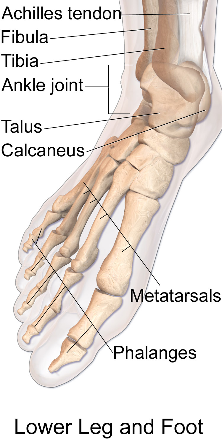


Structure and parts
The talus is traditionally described in three regions: the body, neck and head. The superior surface of the body, the trochlea, fits into the ankle mortise formed by the tibia and fibula and bears much of the joint's articular cartilage. The posterior aspect includes tubercles and a groove for tendons. The inferior surface presents facets that articulate with the calcaneus, and the anterior head connects with the navicular bone.
Articulations and function
Functionally, the talus transmits axial load and allows dorsiflexion, plantarflexion, inversion and eversion through its joints. Major articulations are with the tibia and fibula above, the calcaneus beneath and the navicular in front. Unlike many bones, the talus has no muscular attachments; its stability depends on ligaments, bony fit and surrounding soft tissues.
Blood supply and clinical importance
The talus receives blood from branches of nearby arteries (including branches from posterior and anterior tibial systems and perforating peroneal branches). Its vascular supply is relatively limited, which explains why fractures — particularly of the talar neck — carry a significant risk of avascular necrosis if blood flow is disrupted. Imaging such as X‑ray, CT or MRI are commonly used to evaluate talar injuries.
Injuries, treatment and outcomes
Talar fractures and dislocations most often result from high‑energy trauma, falls or severe ankle twists. Management ranges from immobilization for minor, stable fractures to open reduction and internal fixation for displaced fractures. Complications may include nonunion, post‑traumatic arthritis and avascular necrosis, making early diagnosis and proper reduction important for long‑term function.
Notable facts and resources
- The talus is adjacent to the calcaneus and navicular bones and contributes to the subtalar and talonavicular joints.
- Because most of the talus is covered in articular cartilage, its surface is adapted for joint movement more than for muscle attachment.
- For general references on foot anatomy and clinical approaches, see further reading at anatomy and orthopedics resources and broader clinical summaries at tarsal bone reviews or regional guides at ankle care pages.
Author
AlegsaOnline.com Talus (ankle bone): anatomy, function and clinical significance Leandro Alegsa
URL: https://en.alegsaonline.com/art/96163