
Optic chiasma: anatomy, development, and clinical significance
The optic chiasma is the X-shaped brain structure where optic nerve fibers partially cross; it lies at the base of the brain near the hypothalamus and is crucial for binocular vision and visual field organization.
The optic chiasma, often called the optic chiasm, is the structure at the base of the brain where the two optic nerves meet and exchange some of their nerve fibers. It sits just above the pituitary region and immediately below the hypothalamus; see the nearby brain structures for orientation via related brain anatomy. The optic nerves themselves are cranial nerve II (optic nerve description) and carry visual information from each retina toward the brain.
Image gallery
10 Images
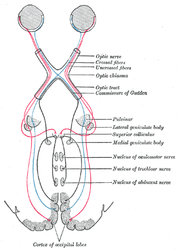


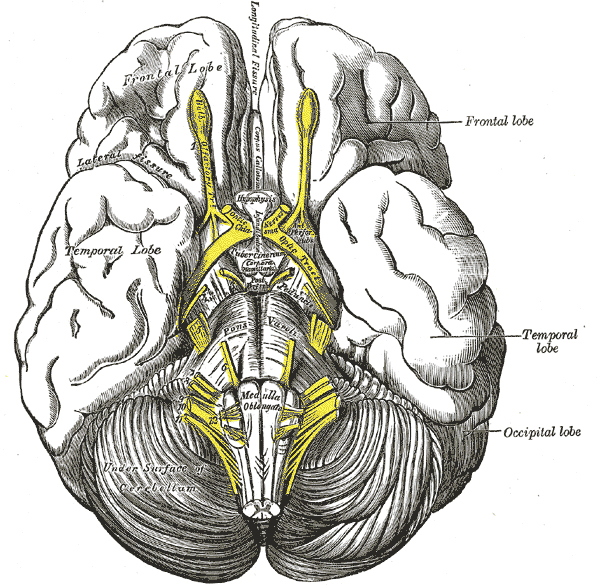


Anatomy and organization
Rather than a complete crossover, the chiasma performs a partial decussation: fibers from the nasal (inner) half of each retina cross to the opposite side, while temporal (outer) retinal fibers remain on the same side. This arrangement reorganizes visual information so that the left cerebral hemisphere receives input from the right visual field and the right hemisphere from the left visual field. The chiasma lies at the inferior surface of the brain near the hypothalamus (hypothalamic region) and is closely related to the pituitary gland and local blood vessels.
Development and name
During embryonic development the optic nerves form as outgrowths of the diencephalon and meet at the midline to create the chiasmal structure. The term "chiasma" derives from the Greek letter chi, reflecting the approximate X-shaped crossing of fibers.
Clinical significance
Because of its compact location, the optic chiasma is vulnerable to compression by nearby lesions, most commonly pituitary adenomas and sellar masses. Compression typically produces characteristic visual field defects, most classically bitemporal (outer) field loss. Other causes of chiasmal dysfunction include inflammatory demyelination, congenital malformations and aneurysms of adjacent vessels. Assessment uses clinical visual-field testing, ophthalmoscopy and neuroimaging.
Notable facts: the partial crossing at the chiasma is essential for binocular integration and depth perception; lesions produce predictable patterns that help localize pathology. For more detailed pathway maps and clinical pictures consult comprehensive neuroanatomy or ophthalmology resources.
Author
AlegsaOnline.com Optic chiasma: anatomy, development, and clinical significance Leandro Alegsa
URL: https://en.alegsaonline.com/art/72881
Sources
- arxiv.org : 1003.1872
- doi.org : 10.1163/157075611X617102
- worldcat.org : 1570-7555