Sebaceous hyperplasia: overview, signs, diagnosis, and treatment
Sebaceous hyperplasia is a benign enlargement of oil glands causing small yellowish papules on the face. This article reviews appearance, causes, diagnosis, management, and when to seek care.
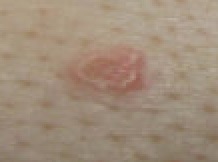
Sebaceous hyperplasia is a common, noncancerous skin condition in which sebaceous (oil) glands become enlarged and form small papules. Lesions are most often found on the forehead, nose, and cheeks of middle-aged and older adults. They usually appear as soft, yellowish or flesh-colored bumps with a central depression or pore-like opening. Many people notice that a small amount of oily material can be expressed from the lesion if gentle pressure is applied, but squeezing is discouraged because it can cause irritation or infection.
Image gallery
7 Images


Clinical features
Typical lesions are:
- Small (few millimeters), dome-shaped papules
- Yellow to skin-colored, sometimes with a central umbilication
- Multiple and clustered, most commonly on the central face
- Generally painless and slow-growing, though they can be cosmetically bothersome
Causes and risk factors
Sebaceous hyperplasia results from enlargement of normal sebaceous glands rather than an infectious process. Contributing factors include aging of the skin, long-term sun exposure, and hormonal influences that affect oil gland function. It is more frequent in middle-aged and older adults. Individuals with altered immune function or those taking some immunosuppressive medications may have a higher occurrence, but the condition is benign in most cases.
Diagnosis and differential diagnosis
Diagnosis is usually clinical, based on appearance and location. Dermoscopy can help identify characteristic features. When a lesion looks atypical, grows rapidly, bleeds, or has unusual features, a skin biopsy is performed to exclude other conditions such as basal cell carcinoma, sebaceous adenoma, sebaceous carcinoma, or milia. Because some malignant tumors can mimic benign glandular growths, practitioners err on the side of biopsy when in doubt.
Treatment and prognosis
Treatment is optional when lesions are asymptomatic. Options for those who want removal for cosmetic reasons or when diagnosis is uncertain include:
- Topical retinoids to reduce gland size over time
- Cryotherapy or electrosurgery to ablate individual lesions
- Laser therapy and photodynamic therapy in selected cases
- Surgical excision for solitary or suspicious growths
If a lesion changes rapidly, bleeds, or becomes painful, seek dermatologic evaluation. For general information and clinical guidance, see professional resources.
Related articles
Author
AlegsaOnline.com Sebaceous hyperplasia: overview, signs, diagnosis, and treatment Leandro Alegsa
URL: https://en.alegsaonline.com/art/88383
Sources
- emedicine.medscape.com : "Sebaceous Hyperplasia: Background, Pathophysiology, Etiology"