Parietal bone
Paired cranial bones forming the sides and roof of the skull. They protect the brain, provide muscle attachments, develop by intramembranous ossification, and join neighboring bones at sutures.
The parietal bones are a pair of large, roughly quadrilateral bones that form much of the superior and lateral aspects of the human cranial vault. In anatomical terminology each is called an os parietale. Together they contribute to the protection of the cerebral hemispheres and provide surfaces for soft-tissue attachments. The name derives from Latin paries, meaning "wall." For general context see skull anatomy.
Image gallery
10 Images


Anatomy and distinguishing features
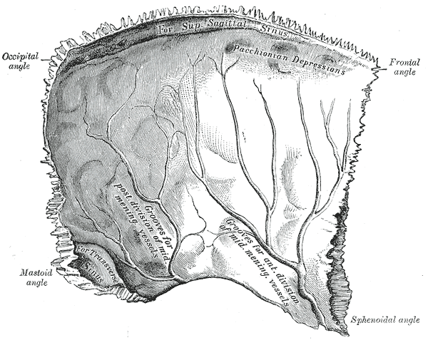
Each parietal bone has an outer (external) and inner (internal) surface, and four borders that articulate with adjacent bones. The main sutures are the sagittal suture between the two parietals, the coronal suture with the frontal bone, the lambdoid suture with the occipital bone and the squamous border toward the temporal bone. Notable surface landmarks include the parietal eminence and occasional parietal foramina that transmit emissary veins. The parietal bones overlie the brain's parietal lobes and may show grooves for meningeal vessels on their inner surfaces.
Development and evolutionary context
Parietal bones form in the cranial vault by intramembranous ossification during fetal and early postnatal growth. They are separated from neighbouring bones by fibrous sutures that allow skull expansion with brain growth; those sutures close gradually across childhood. In evolutionary terms, paired dermal roofing bones such as the parietals are common across vertebrates, and their relative size and shape vary among species.
Function and clinical relevance
Functionally the parietal bones protect cerebral tissue and help maintain the shape of the cranium. They also serve as attachment sites for muscles of mastication and the scalp — for example the temporalis muscle has fibres near the temporal border. Clinically, parietal fractures may accompany head trauma and can transmit force to the underlying brain. Abnormal early fusion of cranial sutures that involve the parietals can alter skull shape and affect intracranial development. For surgical or educational references see cranial bones and clinical skull anatomy.
- Articulations: with frontal, occipital, temporal and sphenoid bones.
- Variations: size and curvature differ between individuals; small foramina may be present.
- Comparative note: paired parietal roofing bones occur across many tetrapods, reflecting a long evolutionary history.
Understanding the parietal bone's form, development and connections is important in anatomy, forensic assessment and neurosurgical planning. Basic diagrams and clinical descriptions are available through standard anatomical references and educational resources cited above.
Author
AlegsaOnline.com Parietal bone Leandro Alegsa
URL: https://en.alegsaonline.com/art/74650