
Blunt trauma: causes, injuries, assessment and management
Blunt trauma (blunt force trauma) is injury from non‑penetrating impacts. This article outlines mechanisms, common injury patterns, clinical assessment, emergency care, imaging, complications, prevention and prognosis.
Overview
Blunt trauma describes physical injury produced by impact with a broad, dull, or firm surface rather than by an object that pierces the body. Energy transferred to tissues during collisions, falls, crush events, or strikes can cause damage beneath intact skin. Common settings include motor vehicle crashes, falls from height, sports injuries, workplace incidents, and assaults. Blunt trauma contrasts with penetrating trauma, in which an object such as a bullet or knife enters the body.
Image gallery
7 Images

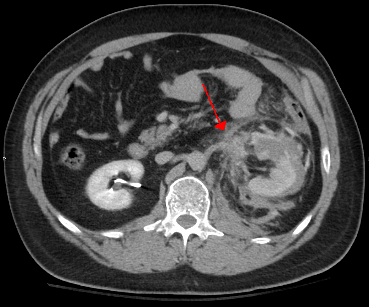


Mechanisms and characteristic injury patterns
Several mechanical processes underlie blunt injuries:
- Compression: direct force squeezes tissues and bones, producing contusions, fractures, or organ rupture.
- Shearing: different tissues move at different speeds, tearing blood vessels or parenchyma.
- Acceleration–deceleration: inertial forces can injure the brain, aortic isthmus, and other mobile organs.
- Crush: prolonged pressure causes soft‑tissue damage, compartment syndrome, and systemic metabolic effects.
Typical anatomic consequences include soft‑tissue bruising and hematoma, closed fractures, pulmonary contusion, splenic or hepatic laceration without external wound, traumatic brain injury, and cardiac contusion.
Clinical assessment and initial management
Evaluation follows standard trauma priorities: airway with cervical spine protection, breathing, and circulation (the ABCs). A primary survey identifies immediately life‑threatening problems; a secondary survey performs a head‑to‑toe examination and documents injuries. Important practical points include immobilizing the cervical spine when indicated, controlling external hemorrhage, and rapidly identifying signs of concealed bleeding such as hypotension, abdominal tenderness, or a declining level of consciousness.
Imaging and monitoring
Diagnostic tools are chosen according to patient stability and suspected injuries. Choices commonly include:
- Plain radiographs for suspected fractures and some chest injuries.
- Focused Assessment with Sonography in Trauma (FAST) to detect free intraperitoneal or pericardial fluid when quick bedside information is needed.
- Computed tomography (CT) for detailed evaluation of the head, chest, abdomen, and pelvis in stable or stabilized patients.
- Continuous monitoring of vital signs, repeated examinations, and serial imaging when injuries may evolve.
Emergency interventions and surgical indications
Treatment aims to stabilize the patient, control hemorrhage, and preserve organ function. Interventions range from observation and supportive care to urgent procedures. Examples include chest tube insertion for pneumothorax or hemothorax, endovascular or open control of major hemorrhage, and laparotomy for uncontrolled intra‑abdominal bleeding. Orthopedic fixation may be required for unstable fractures. Decision making balances the patient’s physiology, injury pattern, and available resources.
Common injury types and special considerations
- Head injury: from minor concussion to severe intracranial hemorrhage; monitoring for delayed deterioration is crucial.
- Thoracic injury: rib fractures, flail chest, pulmonary contusion, cardiac contusion, and great vessel injury.
- Abdominal and pelvic injury: solid‑organ lacerations (spleen, liver), hollow‑viscus injury, and pelvic fractures with hemorrhage.
- Extremity injury: closed fractures, vascular injury, compartment syndrome, and crush‑related muscle damage with systemic effects.
- Crush syndrome: prolonged compression can lead to rhabdomyolysis, hyperkalemia, and renal failure; early fluid resuscitation and monitoring are important.
Special populations
Children and older adults often differ in injury patterns and physiologic reserve. Children may sustain significant internal injury with minimal external signs; older adults commonly have frailty, anticoagulant use, and a higher risk of complications. Pregnant patients require attention to both maternal stabilization and fetal monitoring.
Complications and prognosis
Complications include concealed hemorrhage, organ failure, infection, thromboembolism, long‑term disability from brain or spinal injury, and psychosocial effects. Prognosis depends on the injury severity, promptness and appropriateness of care, comorbidities, and rehabilitation. Early recognition and targeted management improve outcomes.
Prevention and public health
Many causes of blunt trauma are preventable. Measures such as road‑safety laws, seat belts and airbags, fall prevention for older adults, workplace safety practices, and protective sports equipment reduce incidence and severity of injuries.
Forensic and medico‑legal aspects
In forensic contexts, the pattern, distribution, and age of bruises, imprint marks, and fractures help reconstruct events and may distinguish accidental from intentional injury. Documentation, photography, and careful clinical notes are important when medico‑legal issues arise. For additional background see related resources.
Note: This article is an overview of blunt trauma and does not replace professional medical assessment and emergency care.
Author
AlegsaOnline.com Blunt trauma: causes, injuries, assessment and management Leandro Alegsa
URL: https://en.alegsaonline.com/art/12419
Sources
- emedicine.medscape.com : "Forensic Autopsy of Blunt Force Trauma"