Systole
![]()
The title of this article is ambiguous. For other meanings, see Systole (disambiguation).
![]()
This article or subsequent section is not sufficiently supported by evidence (e.g., anecdotal evidence). Information without sufficient evidence may be removed in the near future. Please help Wikipedia by researching the information and adding good supporting evidence.
Systole ([ancient] Greek συστολή - [the] contraction or [the] shortening) is a part of the cardiac cycle. Put simply, it is the tightening and thus blood outflow phase of the heart, in contrast to diastole, the relaxation and thus blood inflow phase. In systole, the blood is squeezed out of the right and left ventricles. Systole describes the pumping action of the heart. It determines the pulse and the pulse amplitude.
The duration of systole remains fairly constant even when the heart rate changes, whereas the duration of diastole varies considerably. Systole is about 300 milliseconds long in adult humans.
The systole of the ventricle is divided into a short mechanical myocardial tension phase and a longer blood outflow phase. Immediately before the tightening phase, the ventricles are filled with blood and the leaflet and semilunar valves are closed. The heart muscle contracts, causing the pressure to rise. In the outflow phase, the pressure in the ventricles exceeds the pressure in the truncus pulmonalis (pulmonary artery) and aorta, the semilunar valves open, and blood flows into the great vessels.
The mechanical discharge phase begins
- in the phonocardiogram or auscultatory with the 1st heart sound
- in echocardiography with the opening of the aortic valve
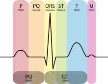
- in the ECG with the R wave
The mechanical discharge phase ends
- in the phonocardiogram or auscultatory with the second heart sound
- in echocardiography with the closure of the aortic valve
- in the ECG with the T-wave
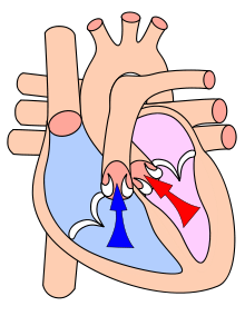
Before systole, the ventricles are dilated and filled with blood, and the atria are contracted. During systole, the two chambers contract simultaneously. The blood is thus pumped into the pulmonary artery and the aorta, from where it flows to the periphery of the body and lungs. At the same time, the atria dilate and fill with blood. To prevent blood from flowing from the ventricles into the atria during systole, their access is closed by the leaflet valves in a valve-like fashion. After systole, the muscle relaxes (diastole). The blood collected in the atria flows through the opening leaflet valves into the ventricles. The leaflet valves are now closed to prevent the blood that was pumped into the aorta and pulmonary artery during systole from flowing back into the ventricles.
The excitability of the heart muscle is suspended during systole (absolute refractoriness).
Note: Systole of the atrium coincides with late diastole of the ventricle.

Schematic representation of an ECG with designations

During systole, blood is pumped into the systemic circulation (red) and the pulmonary circulation (blue).
Disease Patterns
- extrasystole, an extra heartbeat, with different causes
Questions and Answers
Q: What is systole?
A: Systole is the last stage of a heart beat.
Q: When does systole occur?
A: Systole occurs right after diastole.
Q: What happens during systole?
A: During systole, the heart's two ventricles contract (squeeze).
Q: What are the ventricles?
A: The ventricles are the largest parts of the heart, which pump blood to the body and lungs.
Q: What triggers the heart to contract during systole?
A: The heart muscle gets an automatic electrical signal that tells the heart to contract (squeeze).
Q: What is the purpose of systole?
A: The purpose of systole is to make the blood flow out of the heart.
Q: What is diastole?
A: Diastole is the stage of a heart beat when the heart refills with blood.
Search within the encyclopedia