
Staining (Microscopy): Principles, Methods, and Applications
Staining enhances contrast in microscopy by using dyes and labeling methods to reveal structures in cells and tissues for research, diagnostics, and teaching.
Overview
Staining is a set of techniques used in microscopy to increase visual contrast between components of biological specimens. By selectively coloring cells, organelles, or macromolecules, stains make structures easier to detect and interpret under light, fluorescence, or electron microscopes. Staining supports observation of morphology, identification of cell types, and localization of particular chemical groups.
Image gallery
10 Images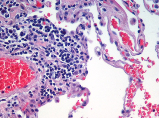


Principles and Categories
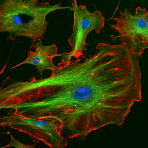
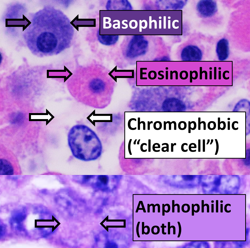
Most stains interact with tissue by chemical affinity, ionic attraction, hydrophobic binding, or specific molecular recognition. Broad categories include general histological stains that color many tissue elements, differential stains that distinguish classes of organisms or cell types, and specific labels such as dyes conjugated to antibodies or nucleic acid probes. Fluorescent stains emit light when excited and are widely used for high-sensitivity localization.
Common Stains and Techniques
- General histology: routine dyes that reveal overall tissue architecture.
- Differential staining: methods that separate groups (for example, bacterial types) by color differences.
- Specialized labeling: stains targeting lipids, carbohydrates, or nucleic acids; enzyme histochemistry; and immunostaining for proteins.
- Fluorescent probes: organic fluorophores and fluorescent proteins used in fluorescence microscopy and live-cell imaging.
Procedure and Types of Preparation
Preparation varies with goals. In vitro staining is applied to fixed, sectioned, or smear preparations; fixation preserves structure and increases stain uptake. In vivo staining involves introducing dyes into living organisms for dynamic studies but is limited by toxicity and permeability. Steps commonly include fixation, embedding, sectioning, staining, washing, and mounting. Controls and standardized timing are important to obtain reproducible contrast.
History and Development
Staining grew alongside microscopy as a practical response to low natural contrast in biological specimens. Early dyes and simple dyesets evolved into modern methods that combine chemistry and molecular biology—antibody-based detection and fluorescent labels greatly expanded specificity. Advances in optics and digital imaging have further increased the utility of staining by allowing multiplexed visualization and quantitative analysis.
Uses, Examples, and Considerations
Staining is central to both research and clinical practice. It is used in biological study and laboratory workflows (biological research) to investigate cell structure, development, and pathology, and in medicine for tissue diagnosis and microbiology (clinical diagnosis). Important considerations include stain specificity, potential artifacts, safety, and compatibility with downstream imaging. Choosing an appropriate technique balances desired contrast, resolution, and preservation of specimen integrity.
Notable Distinctions
Distinguish between nonspecific dyes that broadly tint material and targeted methods that reveal single molecules or activities. Fluorescent and chromogenic stains differ in detection mode and sensitivity. Additionally, some stains are qualitative while others support quantitative image analysis. Proper interpretation requires knowledge of staining chemistry and microscopy limits.
Questions and answers
Q: What is staining used for in microscopy?
A: Staining is used to make cells and tissues easier to see and understand, and to improve contrast in the microscopic image.
Q: What are stains and dyes often used for in biology and medicine?
A: Stains and dyes are often used to highlight structures in biological tissues, often with the aid of different microscopes.
Q: Can staining be done on living tissues?
A: Yes, staining can be done on living tissues (in vivo).
Q: Can staining be done on dead tissues?
A: Yes, staining can be done on dead tissues (in vitro).
Q: Why is staining important in microscopy?
A: Staining is important in microscopy because it enhances visibility and detail of cells and tissues.
Q: What are the benefits of using stains and dyes in microscopy?
A: The benefits of using stains and dyes in microscopy include easier visualization of cells and tissues, improved contrast, and identification of specific structures.
Q: Is staining widely used in the fields of biology and medicine?
A: Yes, staining is a widely used technique in the fields of biology and medicine.
Related articles
Author
AlegsaOnline.com Staining (Microscopy): Principles, Methods, and Applications Leandro Alegsa
URL: https://en.alegsaonline.com/art/93267