Skin: structure, functions, variation, and human significance
Comprehensive overview of skin as an organ and animal covering: its structure, functions, variation among species, health and care, evolutionary background, and cultural uses.
Skin is the external covering of animals that serves as a multifunctional interface between the body and the environment. In vertebrates the integument combines mechanical protection with physiological roles such as temperature regulation, water balance and sensation. Human skin is often described as the body's largest organ; it provides a barrier against infection and physical stress while contributing to identity, social signalling and health.
Image gallery
7 Images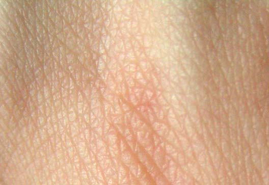


Structure and basic components
Across mammals and many other animals the integument is organized into layers and accessory structures. Typical layers include an outer epidermis of specialized cells, a deeper dermis containing connective tissue, blood vessels and nerves, and an underlying hypodermis of fat and connective tissue. Accessory parts include hair or fur, glands that produce sweat and oils, and pigment cells that determine coloration. These structural elements work together to reduce the entry of microbes such as bacteria and to limit infestations by parasites, while supporting sensory and immune functions.
Primary functions
- Barrier and immune defence: The outer layer resists abrasion and helps prevent invasion by pathogens; it also participates in the immune system's detection of damage.
- Thermoregulation: Skin controls heat loss by blood flow changes, insulation from hair or fat, and evaporation of sweat; the latter process depends on surface evaporation.
- Sensation: Nerve endings in the skin detect touch, temperature and pain, providing critical feedback for behaviour and safety.
- Protection from radiation: Pigmentation and other mechanisms reduce the effects of ultraviolet radiation (UV), and skin participates in vitamin D production.
- Communication and camouflage: Colour, patterns and structures such as hair may signal health, reproductive state or provide concealment.
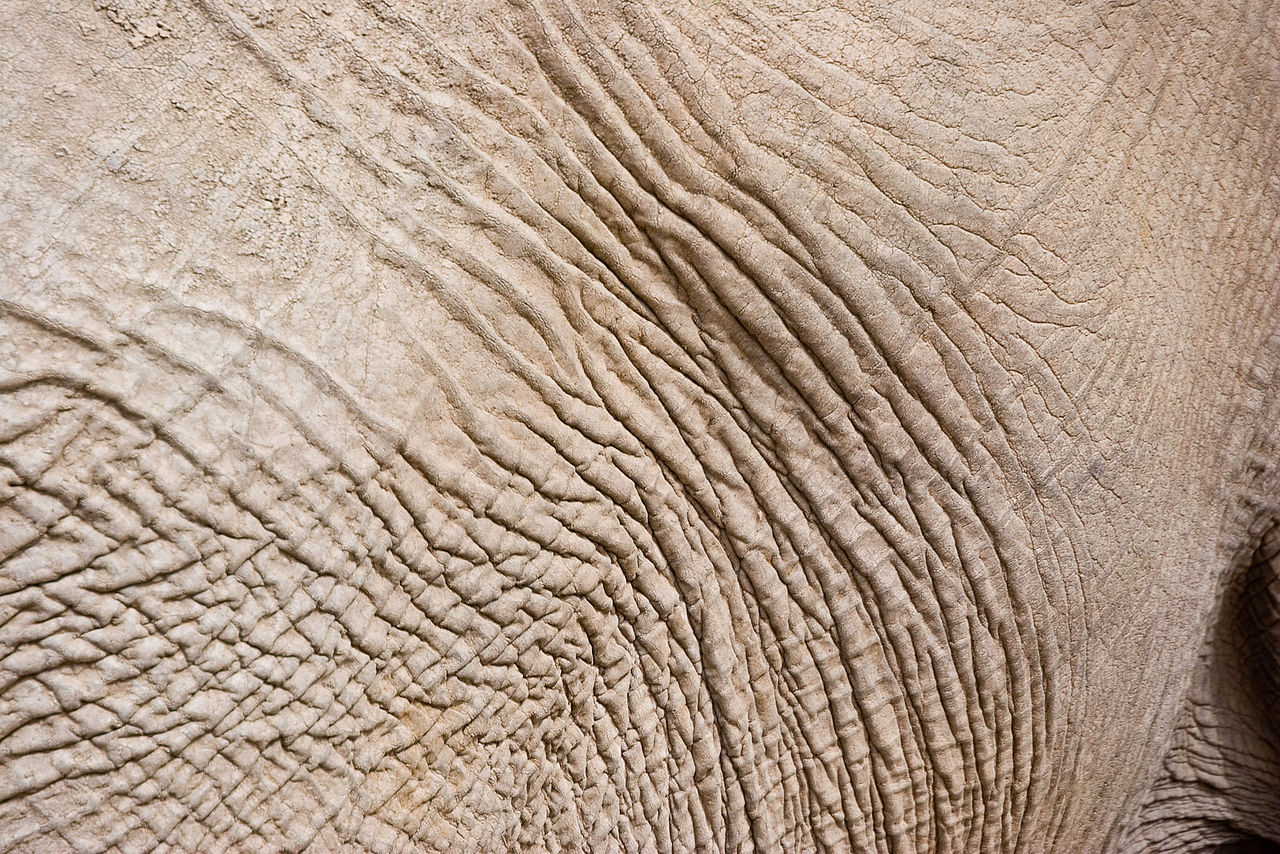
Variation among animals
Different animal groups have evolved distinct coverings and specializations for particular environments. Mammals typically possess hair or fur (mammals; hair, fur) used for insulation and display. Birds are covered in avian feathers (feathers) that aid flight, insulation and signalling. Fish and many reptiles have external scales or scutes (reptiles, including snakes and lizards) that provide armor and reduce water loss. The term scales applies to diverse structures with different developmental origins across groups.
Human skin: variation, culture and care
Human skin colour varies widely around the globe, reflecting differences in melanin production shaped by genetics (genetics) and ancestral exposure to sunlight; social categories such as black people and white people are cultural labels applied to visible variation, but biological pigmentation is continuous. Healthy skin helps prevent infections, supports temperature control and contributes to wellbeing. Routine hygiene and care are important to preserve barrier function and address conditions such as dermatitis, infections or sun damage.
History, evolution and human uses
In evolutionary terms, the development of a keratinized outer layer was a key adaptation for life on land because it reduced desiccation and protected underlying tissues. Over time dermal specializations emerged for insulation, display and mechanical protection. Humans and other cultures have long used animal skins and hides as materials: treated skins become leather, which is fashioned into goods such as shoes, bags and sporting balls. Modern practices and ethics influence how animal hides are sourced and used.
Understanding skin—its biology, diversity and role in health—bridges medicine, ecology and culture. For further reading on immune interactions, pigmentation, coverings in other animal groups and material uses, follow specialist resources and reviews available through scientific and educational portals (microbial risks, immunity, organ overviews).


Etymology
The old Germanic word mhd., ahd. hūt ("skin, integument, epidermis, membranous structure, meninges, fur") belongs to the idg. root [s]keu- extended with t "cover, envelop" and thus means "cover".
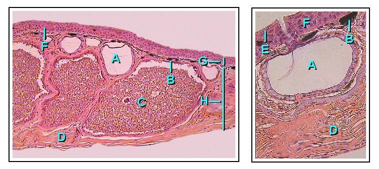
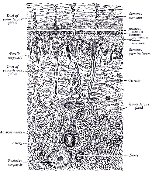
Structure of the human skin
The thickness of human skin is 1.5 to 4 mm. The body surface (skin area) of adult humans is on average 1.73 m². It weighs about 10 to 14 kg.
Layers/components of the skin
The outer skin is divided into three main layers: Epidermis (epidermis), Dermis (dermis, lat. Corium) and Subcutis (hypodermis). The epidermis and dermis together form the cutis.
Epidermis
The epidermis belongs to the epithelial tissues. It is a multilayered keratinizing squamous epithelium that is usually between 0.03 and 0.05 millimeters thick. On the palms of the hands and the soles of the feet, the horny layer is up to several millimeters thick and is colloquially called "callus" (see also horny callus).
The following layers are distinguished from the outside to the inside:
- Horny layer (stratum corneum)
- Luster layer (stratum lucidum) (is only present on the groin skin of the inner sides of the hands and feet)
- Granule cell layer (stratum granulosum)
- Spiny cell layer (stratum spinosum)
- Basal layer (stratum basale)
The prickle cell layer and basal cell layer together form the germinal layer (stratum germinativum).
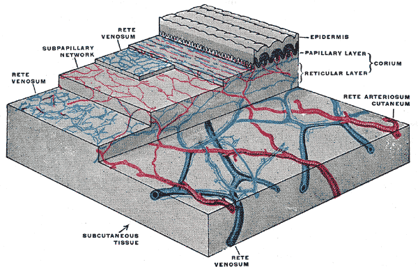
Dermis (dermis, corium)
The dermis consists mainly of connective tissue fibres and serves to nourish and anchor the epidermis. Here the finely capillarized blood vessel system supplies the border zone to the epidermis. The lower dermis contains the smooth muscles and blood vessels that are important for temperature regulation.
The dermis is divided into a stratum papillare (papillary layer, cone layer, papillary body) and a stratum reticulare (reticular layer).
Skin appendages
The skin appendages include various structures such as the scales of reptiles, the feathers of birds, the hair of mammals and other skin-derived structures such as horns, nails, claws and hooves, the substance of which also consists essentially of keratins.
In addition to these structures, skin glands are also included, which open at the epidermis and are anchored in the dermis. In humans, these include sebaceous glands, eccrine sweat glands and scent glands; the mammary gland is a specialized skin gland. The hair-bellows muscle, Musculus arrector pili, which raises a hair, is also an appendage of the skin; contractions of the hair-bellows muscles lead to goose bumps in humans, and in the case of echinoderms they make their hair coat an effective defensive weapon.
Subcutis
The subcutis (or hypodermis) forms the base for the overlying layers of skin and contains the larger blood vessels and nerves for the upper layers of skin, as well as the subcutaneous fat and loose connective tissue. Sensory cells for strong pressure stimuli, for example the lamellar corpuscles, are located in the subcutis.
Surface structure of the skin
If you look at the skin more closely or with a magnifying glass, a fine relief becomes visible. According to this, the skin is divided into two types.
Inguinal skin
Inguinal skin occurs on the fingers, the inside of the hand (palmar) and the sole of the foot (plantar). The epidermis here shows fine papillary lines (skin ridges), which are formed by the dermal papillae being arranged in longitudinal rows. Each dermal ridge is underlain by two rows of papillary bodies. The dermal ridges form an individual pattern of various geometric figures (whorls, arches, loops, double loops). These patterns are used forensically in dactyloscopy (fingerprint recognition) as a form of biometric data. The skin of the groin does not contain any skin appendages, except for many sweat glands.
Field skin
Field skin covers the remaining skin areas. Here the surface shows rhombic fields delimited by fine furrows (areolae cutaneae). The furrows are formed on the epidermal areas that are free of papillae and disappear when the skin is under greater tension. They serve as reserve folds because the epidermis is less stretchable than the dermis. The size of the dermal fields varies with the region of the body. The field skin contains the skin appendages and is less than 0.1 mm thick. It is thinnest in the region of the eye and the genital organs.


Related articles
Author
AlegsaOnline.com Skin: structure, functions, variation, and human significance Leandro Alegsa
URL: https://en.alegsaonline.com/art/90932