Radiography: principles, equipment, clinical uses and safety
Radiography is the use of X‑rays to produce projection images of internal structures. This article explains how radiography works, common equipment, clinical applications, safety considerations and related techniques.
Overview
Radiography refers to medical imaging produced by passing X‑rays through the body to form a two‑dimensional projection image of internal structures. It is one of the oldest and most widely used diagnostic imaging methods. Radiographs help clinicians detect fractures, lung disease, certain abdominal conditions and many other problems quickly and inexpensively. The technique relies on differences in how tissues absorb or attenuate high‑energy electromagnetic radiation compared with visible light and other parts of the spectrum.
Image gallery
10 Images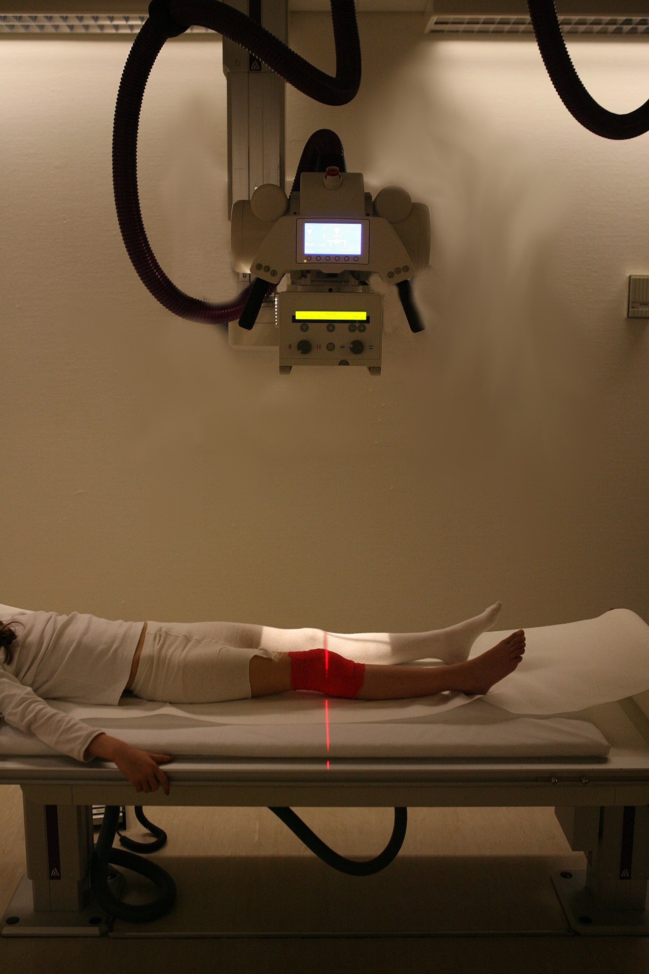


How radiography works
An X‑ray tube generates a controlled beam of X‑rays, a form of high‑energy electromagnetic radiation. When this beam passes through the body, different tissues absorb different proportions of the radiation depending on their density and composition: bone and metal absorb more, soft tissues and air absorb less. The pattern of transmitted X‑rays is captured behind the patient by a detector to form an image. Radiographs are therefore projection images in which structures along the beam are superimposed.
Equipment and image formation
Typical radiography systems have several basic components: an X‑ray source, a collimator to shape the beam, a patient support or table, and a detector. Detectors are either conventional photographic film or modern digital systems such as flat‑panel detectors or computed radiography plates. Digital detectors offer faster processing, manipulation of image contrast, and easier storage and transmission.
- X‑ray tube: produces the radiation and controls beam energy and intensity.
- Collimation and grids: reduce scatter and improve image contrast.
- Detectors: from photographic film and digital detectors to specialized sensors for dental or mammographic imaging.
History and development
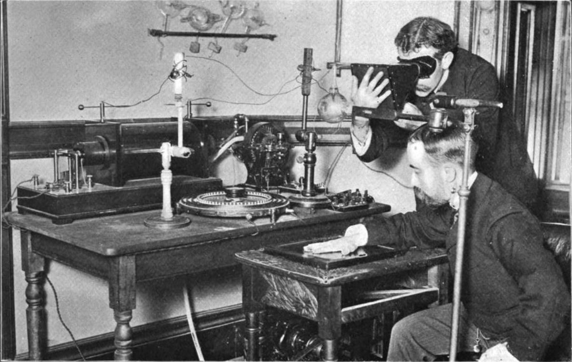
Radiography emerged soon after Wilhelm Röntgen’s discovery of X‑rays in 1895. Early photographic plates recorded the first images of bones and metal objects inside the body. Over the 20th century, improvements in tube design, radiation safety, image intensification and electronic detectors transformed radiography into a core clinical tool. Digital technology has progressively replaced film, enabling lower doses in some studies and greater flexibility in image handling.
Clinical uses and examples
Common radiographic studies include chest radiographs for evaluating lungs and heart, skeletal radiographs for fractures and joint disease, abdominal imaging for obstruction or perforation, dental X‑rays for teeth and jaws, and mammography for breast screening. Radiographs are frequently used as first‑line tests because they are rapid, widely available and relatively inexpensive. Contrast agents are sometimes introduced to outline hollow organs or blood vessels and improve diagnostic detail.
Safety, dose and limitations
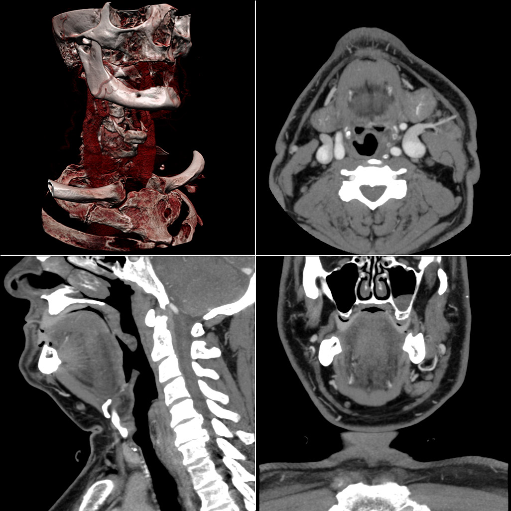
Because X‑rays are ionizing radiation, radiography involves exposure that should be kept as low as reasonably achievable (ALARA). Clinical teams balance diagnostic benefit against potential risk and select alternative modalities—such as ultrasound or MRI—when appropriate. Radiography provides projection images, so overlapping structures can obscure findings; when three‑dimensional detail or cross‑sectional imaging is needed, computed tomography (CT) or other modalities may be preferred.
Related techniques and distinctions
Radiography is distinct from but related to several other imaging methods. Fluoroscopy uses continuous X‑ray beams for real‑time imaging during procedures. CT uses multiple X‑ray measurements from different angles and computational reconstruction to create cross‑sectional images rather than single projection radiographs. For more on the physical principles and the broader electromagnetic spectrum see electromagnetic radiation. Additional references on X‑ray physics and clinical protocols can be found through educational resources and professional guidelines.
For technical details on X‑ray generation and detector types see X‑rays and for information on detector technology consult sources about photographic film and digital detectors. For general comparisons between modalities, including non‑ionizing options such as ultrasound and MRI, see visible light and imaging contrast.
Questions and answers
Q: What is radiography?
A: Radiography is the use of X-rays to see what's happening to parts of the body.
Q: How is radiography done?
A: Radiography is done through an imaging technique that uses electromagnetic radiation other than light, usually X-rays. To create the image, a beam of X-rays is produced by an X-ray machine and projected toward the object.
Q: What happens when X-rays are projected toward an object in radiography?
A: When X-rays are projected toward an object in radiography, a certain amount of X-ray is absorbed by the object, which is dependent on the density and composition of that object. The X-rays that pass through the object are captured behind the object by a detector (either photographic film or a digital detector).
Q: What type of representation does the detector give in radiography?
A: The detector gives a superimposed 2D representation of the object's internal structures in radiography.
Q: Can radiography be used to see the internal structures of objects other than the human body?
A: Yes, radiography can be used to see the internal structures of objects other than the human body.
Q: Is radiography a safe imaging technique?
A: Radiography is a safe imaging technique if used correctly with appropriate safety precautions. The amount of X-ray used should be as low as possible to achieve the desired imaging result to prevent unnecessary exposure to radiation.
Q: What are some common types of medical procedures that use radiography?
A: Some common types of medical procedures that use radiography include X-rays to view bones or teeth, mammography to detect breast cancer, and CT scans to view internal organs and tissues.
Related articles
Author
AlegsaOnline.com Radiography: principles, equipment, clinical uses and safety Leandro Alegsa
URL: https://en.alegsaonline.com/art/80772