
Radiation therapy: principles, techniques, and clinical uses
Radiation therapy uses ionizing radiation to treat cancer and some non-malignant conditions. This article explains how it works, common techniques, clinical roles, planning and safety, and historical development.
Radiation therapy is a medical treatment that uses directed radiation to damage or destroy abnormal cells. It is most commonly applied to cancer, where it can be used with curative, adjuvant, neoadjuvant, or palliative intent. Practitioners typically treat a defined mass or area, such as a visible tumor, adjacent tissues thought to harbor microscopic disease, or regional lymph nodes. Modern practice balances the goal of delivering a high enough dose to control disease against the need to preserve surrounding healthy tissue and maintain quality of life.
Image gallery
8 Images



How radiation therapy works
The therapeutic effect of treatment relies on the biological impact of ionizing radiation on cells. At clinically relevant doses, radiation damages cellular components, most importantly the DNA, causing breaks that interfere with replication and can lead to cell death. Because malignant cells often have impaired DNA repair and more rapid division than normal cells, they are relatively more susceptible to these effects. Radiation oncologists exploit this differential sensitivity through dose fractionation — delivering the total dose in multiple smaller sessions — allowing normal tissues time to repair while maximizing tumor control.
Major techniques and delivery methods
- External beam radiation therapy (EBRT): The most common approach, using a beam of radiation directed from outside the body. Sources include X-ray machines and linear particle accelerators. Advanced EBRT techniques such as intensity‑modulated radiation therapy (IMRT), volumetric modulated arc therapy (VMAT), and image‑guided radiation therapy (IGRT) shape dose distributions tightly around the target.
- Stereotactic radiosurgery and stereotactic body radiotherapy (SRS/SBRT): High-precision methods that deliver large doses in few sessions to small targets, often in the brain or lung, with steep falloff to spare adjacent tissue.
- Particle therapy: Uses charged particles such as protons or heavier ions. Because of their distinct depth-dose characteristics, particles can reduce dose beyond the tumor and limit exposure to normal structures.
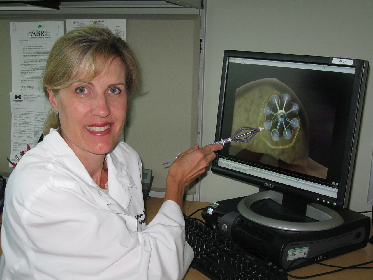
- Brachytherapy: Placement of sealed radioactive sources inside or next to the treatment area. This approach is commonly used in cancers of the breast, prostate and other pelvic or gynecologic sites, delivering high local dose while minimizing distant exposure.
- Total body irradiation (TBI): A whole‑body technique used primarily as part of conditioning before bone marrow or stem cell transplantation.
Indications, combinations, and examples of use
Radiation therapy is adaptable and appears across many clinical scenarios. It may be curative for localized disease, adjuvant after surgery to reduce recurrence risk, or palliative to relieve symptoms such as pain or bleeding. Oncologists commonly combine radiation with other modalities: concurrent or sequential chemotherapy, surgery, hormone therapy, and the growing field of immunotherapy. Patterns of care depend on tumor type, stage, anatomic location, and patient factors. For example, brachytherapy plays a central role in certain prostate and gynecologic cancers, while SBRT is increasingly used for small lung tumors.
Non-malignant applications and considerations
Although most commonly used for oncology, radiation can treat select benign conditions. Indications sometimes include painful joint disorders, prevention of problematic scar tissue such as keloid formation after surgery, and certain vascular or neurologic conditions. Because radiation carries a small long-term risk of inducing secondary malignancy and can cause cumulative tissue damage, its use in non-malignant disease is cautious and limited to situations where benefits clearly outweigh risks.
Treatment planning, targets, and safety
Radiation treatment begins with a careful planning phase. Imaging studies used for planning and verification include CT, MRI, and nuclear medicine techniques; these diagnostic tools are part of the broader field of diagnosis and image guidance. Specialists define a target volume that includes the visible tumor plus margins for microscopic disease and motion; regional lymph nodes may be included when there is a risk of spread. The specialty that prescribes and oversees this therapy is radiation oncology, distinct from radiology, which focuses on imaging. Rigorous quality assurance, careful dose calculations, and measures to account for tumor motion during breathing are essential to minimize unintended exposure. Acute side effects (skin irritation, fatigue) and late effects (fibrosis, organ dysfunction) are monitored and managed by multidisciplinary teams.
History and notable developments
The therapeutic use of radiation has evolved since the early 20th century, progressing from radium sources and rudimentary X‑ray techniques to modern accelerator-based machines and sophisticated planning systems. The introduction of linear accelerators and advances in imaging enabled conformal dose delivery. Research into particle therapy and biologically guided planning continues to refine how treatments are tailored to individual tumors and patients. Innovations aim to improve tumor control while reducing toxicity through better targeting, motion management, and combined modality approaches that exploit radiobiology.
Important distinctions for patients and clinicians include the intent of treatment (curative versus palliative), the selection of modality based on tumor biology and location, and the balance between efficacy and side effects. Patients considering radiation therapy usually consult a multidisciplinary team and a board‑certified radiation oncologist to review expected benefits, likely side effects, and alternatives. For additional technical or educational resources see links on beam sources and accelerators such as particle accelerators, or general references about tumor control, radiation safety, and specific site‑directed strategies for breast, prostate and other organs. For further context on imaging and diagnosis, consult materials indexed under diagnostic imaging and professional guidelines available through specialty organizations.
Application against cancer
Malignant tumors are very often irradiated; often also in combination with other treatment procedures such as surgery and chemotherapy. About every second cancer patient receives one or more radiation therapies. Palliative radiotherapy, for example of bone metastases, and curative treatment, i.e. treatment intended to cure cancer, are about equally common. Neoadjuvant radiotherapy is intended to reduce the size of the tumour for subsequent surgery; adjuvant radiotherapy is intended to secure the result of a previous operation and destroy microscopic tumour nests. Oncological treatment always follows the "log cell kill" principle. Radiotherapy with the intention of healing is designed to destroy the tumour, which often consists of 100 billion cells, down to the last cell. Since individual tumour cells can no longer be detected, the actual success of the treatment only becomes apparent in the months and years that follow. If a tumour forms again at the same site within the follow-up period, it must be assumed that there has been a recurrence.
The treatment decision is based, among other things, on the question of the extent to which the localisation of the tumour to be treated is suitable for radiotherapeutic intervention. Not all tumors are more radiosensitive than the normal tissue surrounding them. One of the causes of lower radiation sensitivity is oxygen deficiency (hypoxia) in the tumor tissue. By means of a combination of fractionation and irradiation technique that is optimally adapted to the tumor biology and the surrounding risk organs, it is now possible to successfully treat even problematically localized and relatively radiation-insensitive tumors. An optimal irradiation technique delimits the tumor region supplied with dose by the steepest possible dose fall-off to normal tissue. Various therapy concepts additionally attempt to increase the radiation sensitivity of tumors with the aid of so-called radiosensitizers (radiation sensitizers).
The curative effect requires a total dose of 20 to 80 Gray, depending on the tumor type and fractionation, administered in one session or spread over several weeks, depending on the treatment regimen. Symptom-relieving treatments for incurable patients can be shorter; for example, bone foci can be treated with a single 8 Gy of pain relief.
With modern radiotherapy procedures, a large number of tumor diseases can be cured today in stage-dependent combination with surgery and chemotherapy, even in advanced stages. Averaged over all tumor types and stages, the chance of cure is about 50 %. Individual tumours such as Hodgkin's disease and seminoma of the testis can almost always be cured. The most common indications for radiotherapy are currently prostate cancer, adjuvant after breast cancer surgery, and for rectal cancer. A particular advantage is the fact that radiotherapy can preserve organs even in situations where the disease is already relatively advanced. The combination of radiotherapy with chemotherapy in the case of cancer of the larynx can be mentioned here in particular. In other tumor diseases, such as prostate carcinoma, surgical procedures and radiotherapeutic procedures compete with each other and can have comparable results. Here, it is the task of the consulting physician to explain to the patient all the advantages and disadvantages of the respective procedures. In this context, it is desirable to have certified tumour centres in which all specialist disciplines are represented and which thus enable the patient to receive comprehensive advice.
Very rarely, an abscopal effect can occur, in which tumor regression up to complete remission is recorded even at sites that were not irradiated. This effect was first described in 1953 and has so far only been reported in isolated cases, such as leukaemia, lymphoma, renal cell carcinoma and malignant melanoma.


Application against benign diseases
→ Main article: Radiotherapy for benign diseases
Numerous chronic inflammatory and degenerative diseases such as heel spurs, tennis elbow, shoulder pain, arthroses of the various joints, vertebral body hemangiomas, induratio penis plastica and others can be treated with radiation therapy. This so-called stimulus radiation far below the tissue-killing dose is free of side effects except for the stochastic risk. Response rates are in the range of 50 to 70 %. The total doses used are in the range of 1 to 20 Gray, which is much lower than the doses that must be used in cancer therapy (20 to 80 Gy). Low-dose radiation reduces the activity of leukocytes and connective tissue precursor cells and decreases the release of cytokines; thus, it inhibits acute and chronic inflammatory processes. Direct inhibition of pain receptors is also suspected. Shoulder pain and heel spurs in particular are readily amenable to radiation therapy. In Germany, about 37,000 patients with non-malignant diseases are irradiated per year, with an increasing tendency.
Related articles
Author
AlegsaOnline.com Radiation therapy: principles, techniques, and clinical uses Leandro Alegsa
URL: https://en.alegsaonline.com/art/80741