
Protein structure and folding: architecture, dynamics, and methods of study
Overview of how proteins are built and folded, levels of structural organization, forces that shape them, experimental and computational methods, biological roles, and examples of conformational change.
Protein structure describes the three-dimensional arrangement of a single protein molecule and how its parts are organized in space. Proteins are central biological macromolecules found across all organisms; they are linear polymers made during translation from a sequence of about 20 standard amino acids. The sequence of amino acids (the primary structure) determines the ways a polypeptide will fold and what shapes and interactions it can form.
Image gallery
6 Images

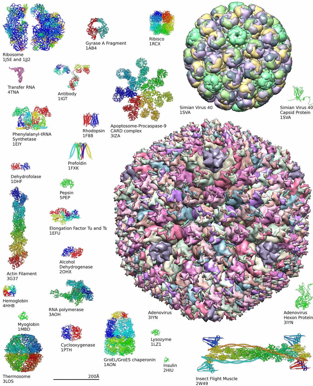


Levels of structure and common elements
Structural biology commonly describes protein architecture at several levels. Primary structure is the amino acid sequence. Secondary structure refers to local regular conformations, such as alpha helices and beta sheets, stabilized by backbone hydrogen bonds. Tertiary structure is the overall three-dimensional fold of a single chain, shaped by side-chain packing and a balance of forces; quaternary structure arises when multiple chains assemble into a functional complex.
Forces driving folding and stability
Folding is not based solely on rigid chemical bonds like covalent links (except for disulfide bridges); rather it is driven by many weaker interactions: hydrogen bonding, electrostatic interactions, van der Waals forces, hydrophobic effect and sometimes metal coordination. The native folded state is usually the conformation with a low free energy under physiological conditions. Folding can be assisted in cells by molecular chaperones and can fail, producing misfolded species that are associated with disease.
Techniques to determine and analyze structure
Experimental methods reveal protein structures at atomic or near-atomic detail. Classical techniques include X-ray crystallography and NMR spectroscopy, while surface-sensitive methods such as dual polarisation interferometry complement those approaches. Cryo-electron microscopy and mass spectrometry-based methods are also widely used. Computational prediction and modeling have advanced rapidly, providing structural hypotheses that guide experiments.
Dynamics, conformations and function
Proteins are dynamic molecules that can switch between different conformations as part of their function. An enzyme, for example, will often adopt distinct conformations when it binds a substrate or a cofactor; these changes can be small local rearrangements or large domain movements. Such conformational flexibility underlies mechanisms like allosteric regulation, induced fit, and mechanical work performed by motor proteins.
Importance, examples and notable distinctions
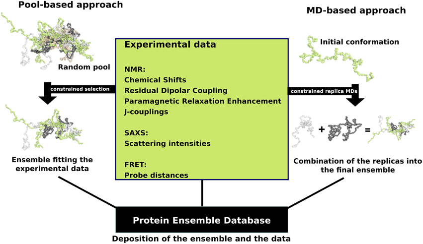
Understanding structure is essential to explain how proteins recognize partners, catalyze reactions, and form cellular machines. Structural knowledge guides drug design, protein engineering and interpretation of genetic variation. Some proteins are intrinsically disordered and function without a single fixed fold, illustrating that biological activity can arise from both ordered structures and flexible ensembles. Errors in folding or assembly are implicated in conditions such as amyloid diseases, highlighting the health relevance of protein structural biology.
- Levels of organization: primary, secondary, tertiary, quaternary.
- Key interactions: hydrogen bonds, hydrophobic effect, ionic interactions.
- Common methods: X-ray crystallography, NMR spectroscopy, dual polarisation interferometry, cryo-EM, computational modeling.
- Functional themes: conformational change, allostery, assembly, disorder.
For further reading on experimental protocols, computational tools and case studies in structural biology, consult specialized texts and databases that catalog solved structures and sequence-structure relationships. Representative resources are available through scientific repositories and institutional collections (proteins, macromolecules, organisms references) and methodological overviews (X-ray, NMR, DPI) that explain experimental design and interpretation.
Questions and answers
Q: What is protein structure?
A: Protein structure refers to how protein molecules are organized and the shape they assume, which influences their function.
Q: Why are proteins important in biology?
A: Proteins are vital biological macromolecules found in all living organisms. They perform a wide range of biological functions, such as building and repairing tissues, transporting molecules, and regulating cellular processes.
Q: How are proteins formed?
A: Proteins are made up of 20 different amino acid polymers, which are created through RNA translation.
Q: What determines a protein's shape?
A: Proteins fold into specific shapes through weaker forces such as hydrogen bonds, rather than chemical bonds.
Q: How can biophysics be used to study protein structure?
A: Biophysics employs various techniques such as X-ray crystallography, NMR spectroscopy, and dual polarisation interferometry to understand the three-dimensional structure of proteins.
Q: How do proteins switch from one shape to another?
A: Proteins may switch between different shapes or conformations as they perform their functions in the body.
Q: What are conformations in relation to proteins?
A: Conformations refer to the different shapes or alternative states that the same protein can assume. For example, an enzyme may have two conformations: one with its co-enzyme and one without.
Related articles
Author
AlegsaOnline.com Protein structure and folding: architecture, dynamics, and methods of study Leandro Alegsa
URL: https://en.alegsaonline.com/art/79517
Sources
- ncbi.nlm.nih.gov : "Protein length in eukaryotic and prokaryotic proteomes"
- doi.org : 10.1093/nar/gki615
- pubmed.ncbi.nlm.nih.gov : 15951512
- ncbi.nlm.nih.gov : "The structure of proteins; two hydrogen-bonded helical configurations of the polypeptide chain"
- doi.org : 10.1073/pnas.37.4.205
- pubmed.ncbi.nlm.nih.gov : 14816373
- ncbi.nlm.nih.gov : ncbi.nlm.nih.gov/pmc/articles/PMC1173893/
- pubmed.ncbi.nlm.nih.gov : 2649166
- doi.org : 10.1046/j.1432-1327.2002.02797.x
- pubmed.ncbi.nlm.nih.gov : 11895428