Prenatal testing
![]()
This article deals with specific prenatal diagnostic procedures. For other preventive measures, see prenatal care.
The term prenatal diagnosis or prenatal diagnostics (abbreviation PND; composed of Latin prae "before" and natal "birth", see prenatal, as well as diagnostics) refers to examinations of fetuses and pregnant women (see also early detection of diseases).
Common methods are, on the one hand, the non-invasive examinations performed only from outside the body, such as ultrasound examination (sonography), which includes nuchal translucency measurement, nasal bone measurement (in the 12th to 14th week of pregnancy), fetometry, fine ultrasound, Doppler sonography, 3D ultrasound and 4D ultrasound. The examinations of hormone concentrations in the maternal blood (serological examinations) such as the triple test, the quadruple test and, as a combination of both methods, the first trimester screening (ETS, an examination in the first trimester of pregnancy) and the integrated screening (combined examination of biochemical serum parameters to determine the risk of fetal chromosomal and occlusion disorders) also belong to the non-invasive methods. Non-invasive diagnostics also include the prenatal paternity test, which is possible as early as the 9th week of pregnancy and in which fetal DNA is isolated and analysed using a blood sample from the mother. Invasive prenatal diagnostic tests, i.e. those performed inside the body, include chorionic villus sampling (CVS), amniocentesis (AC) and umbilical cord puncture. An earlier method of prenatal diagnostics was amnioscopy.

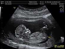
Sonographic image of a fetus at 12 weeks of gestation
History
| Investigations in reproductive medicine |
| Prefertilisation diagnostics:
Preimplantation genetic diagnosis:
Prenatal diagnostics: |
The first amniocenteses for prenatal diagnostics were performed in 1930 by Thomas Menees and others. The foundation of modern prenatal diagnostics was laid by the British obstetrician Ian Donald in 1958 with the first sonographic imaging of an unborn child. The technical advancement of ultrasound equipment enabled a high resolution of detail and thus the detection of structural malformations of fetal organs with a high diagnostic certainty. Thus, the diagnosis of abdominal wall hernias, diaphragmatic hernias, displacements of the heart axis, malformation of organs such as the lungs or kidneys, cystic kidneys, malformations of the extremities, obstructions in the gastrointestinal tract, etc. became possible.
Non-invasive ultrasound technology was extended by the development of invasive techniques. Thus, in 1966, Steele and Breg demonstrated the possibility of sampling and chromosomal examination of fetal cells contained in the amniotic fluid (amniocentesis) during the second trimester of pregnancy. This was followed in the early 1980s by the publication of chorionic villus sampling. In this procedure, cells are taken from the villi of the egg skin (chorion) during the first trimester of pregnancy, which later forms the placenta. These cells are cultured and subjected to genetic analysis. Later, with early amniocentesis, puncture of fetal vessels and fetal organs, and collection of blood from the umbilical cord, further measures for the collection of fetal cells followed.
More recently (as of 2006), fetal MRI has also become increasingly available as a non-invasive diagnostic method. The basis for referral is often a suspected sonographic diagnosis. The clarification of rare syndromes is possible by means of MRI with a high diagnostic certainty.
Up to now, invasive examination procedures have been burdened with different risks that depend on various factors, e.g. that of a miscarriage. As a result, in addition to the basic need of prospective parents worldwide to have a physically and cognitively healthy, non-disabled child, and the desire to know about the child's state of health, the trade-off between the risks of the examination and the likelihood of a particular disability usually plays an important role in the decision to use an invasive examination.
However, this can have far-reaching social and legal consequences: Those doctors who advise against an invasive examination such as amniocentesis or chorionic villus sampling sometimes find themselves exposed to claims for damages in the event of the birth of a child with a disability detectable by the examination. This is one of the reasons why a nationwide offer of prenatal examinations with a high diagnostic certainty has been established in Germany in the meantime, which sometimes gives the impression of "avoidability" of children with disabilities due to the meanwhile quite high degree of awareness of different examination procedures.
As risk-free, albeit non-diagnostic examinations, addiction tests such as the double test, triple test and nuchal translucency measurement as part of first trimester screening are currently popular. They provide indications of a possible chromosomal abnormality or of certain physical malformations, e.g. in the area of the spinal canal, the abdominal wall and the kidneys. The extent to which these procedures should be used across the board in Germany as part of maternity screening is still the subject of controversial ethical and health policy discussions.
Since the early 1990s, intensive research has been conducted worldwide into non-invasive examination methods (NIPT), in which the genetic material of the fetus can be extracted from cells in the blood of the pregnant woman and examined for genetic abnormalities without risk, in order to avoid the use of invasive procedures with a risk of miscarriage for chromosome extraction. According to Hepp, this procedure was in clinical trials in 1999.
According to researchers at Stanford University in California, they succeeded in 2008 in enriching isolated fetal cells present in maternal blood, subjecting them to DNA analysis and thus making many invasive examinations carried out for this purpose superfluous. The NIPT procedure was successful in detecting 12 different chromosomal disorders.
Application Risks
Non-invasive tests
→ Main article: Non-invasive prenatal test
In the case of non-invasive examinations (= examinations that do not penetrate the pregnant woman's body), such as an examination of maternal blood (NIPT) and also in the case of an ultrasound examination such as nuchal translucency measurement, there are no risks for the unborn child and its mother according to the current state of knowledge (2014).
Invasive examinations
In various medical textbooks with research status 2000, 2005 and 2007, approx. 1 % intervention-related risk of abortion is assumed for amniocentesis (amniocentesis) and for chorionic villus sampling.
According to recent studies, invasive (invading the pregnant woman's body) diagnostic procedures such as amniocentesis, chorionic villus sampling, or umbilical cord puncture pose little risk to the fetus.
For example, in 2006, a study examined 35,003 unselected pregnant patients from the normal population. It was found that the risk of miscarriage in patients who did not have amniocentesis was 0.94%, whereas the risk of miscarriage in the women in the study group increased to only 1.0%. Amniocentesis showed no statistically significant effect on the risk of miscarriage in this study.
Another study from 2006, which evaluated 9,886 chorionic villus sampling and 39,893 amniocentesis, shows that there is no significant difference in risk between the two screening methods.
Psychological stress
Until a conclusive test result is available, there is sometimes a restricted mother-child relationship due to the socially and personally at least latently assessed basis as "pregnancy on trial". In some cases, an emotionally strong bond is only allowed when the results are unremarkable, which means that the option of terminating the pregnancy in the case of abnormal results is also kept as emotionally open as possible. Sometimes the time up to this point is psychologically difficult for the pregnant woman; "most feelings of guilt arise from the feeling of having put the child up for disposal", since the health of the fetus is regarded as the decisive criterion for its acceptance or rejection. On the other hand, an unremarkable finding helps to reassure parents. Expectant parents are advised to seek counselling before making use of prenatal diagnostics. Especially in the case of family history, it is advisable to visit a genetic counselling centre.

Search within the encyclopedia