Axilla (armpit): anatomy, function and clinical importance
The axilla, or armpit, is the pyramid-shaped area beneath the shoulder that contains vessels, nerves, lymph nodes, sweat glands and fat; important in clinical examination, thermometry and surgery.
The axilla, commonly called the armpit or underarm and historically known as the oxter, is the pyramidal region on the human body beneath the shoulder. It occupies the space below the point where the arm meets the torso, adjacent to the shoulder joint and the shoulder girdle. This cavity forms a passage between the neck and the upper limb and is lined by skin, subcutaneous tissue and fascia.
Image gallery
10 Images


Anatomy and contents
The axilla has identifiable boundaries and contains several important structures. Its walls are formed by muscles and bones, its apex opens toward the neck and its base is the skin-covered hollow felt beneath the arm. Major contents include:
- Neurovascular bundle: the axillary artery and vein and branches of the brachial plexus that supply the upper limb.
- Lymphatic tissue: multiple axillary lymph nodes grouped in levels that drain the arm and much of the breast and chest wall.
- Soft tissues: adipose tissue, connective tissue and sweat (apocrine and eccrine) glands, plus hair follicles and skin.
These elements make the axilla both a protective corridor for vital vessels and nerves and a site where disease processes may concentrate.
Clinical significance
The axilla is important in medicine for several reasons. It is one of the traditional sites where a medical thermometer can be placed to estimate body temperature, although axillary readings are generally lower than core measurements. Other common temperature measurement sites include the rectum, the mouth and the ear canal. Palpation of axillary lymph nodes is part of routine physical examinations because enlargement can signify localized infection, systemic disease or metastatic spread from malignancies such as breast cancer. Surgical procedures may involve the axilla for biopsy or removal of lymph nodes (sentinel node biopsy or axillary dissection).
Dermatologic and dermatologic-surgical conditions that affect the axilla include excessive sweating (hyperhidrosis), chronic inflammatory follicular disease, and hidradenitis suppurativa. The region's anatomy also influences the placement of deodorants, topical medications and the approach to regional anesthesia.
Development, variation and notable points
During development the axillary space forms as the limb bud grows away from the trunk; anatomical variation is common in the exact course of small vessels and nerves. Clinicians and anatomists emphasize the axilla because it is a crossroads for clinical signs: skin changes, palpable nodes, or abnormal temperature readings can all provide diagnostic clues. When measuring temperature or evaluating lymph nodes, context and technique matter—axillary findings are interpreted alongside other signs and diagnostic tests.
In summary, the axilla is a small but complex region that plays a key role in limb vascularization and innervation, immune surveillance via lymph nodes, thermometry and surface physiology. Its accessibility makes it a frequent focus in both basic examination and specific surgical or medical interventions.


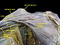
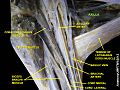
Anatomy of the axilla
axillary fossa
When the upper arm is abducted (spread), the axillary fossa forms a four-sided pyramid, the spatial extent of which is approximately as follows:
| Base: | axillary fascia |
| ventral (anterior) wall: | Fascia clavipectoralis, Musculus pectoralis major, Musculus pectoralis minor |
| dorsal (back) wall: | Musculus subscapularis, Musculus latissimus dorsi, Musculus teres major |
| medial (towards the middle of the body) wall: | anterior serratus |
| lateral (outer) edges: | anterior and posterior axillary folds (formed by the pectoralis major and latissimus dorsi muscles) |
| cranial (upper) wall: | Shoulder joint (articulatio humeri), proximal end of humerus, coracobrachialis muscle and caput breve of biceps brachii muscle |
| Tip: |
· 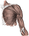
Superficial muscle layer of thorax and upper arm, right side viewed dorsally
· 
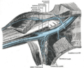
The branches of the brachialartery
· 
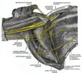
The veins in the right axilla, seen ventrally
· 
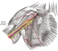
The right brachial plexus (the infraclavicular part), view from the front below
· ![]()
The left side of the chest
· 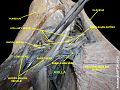
Axilla
· 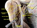
Axilla
· 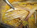
Axilla
· 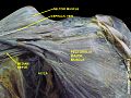
Axilla
· 
Axilla
· 
Axilla
· 
Axilla
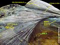
Axle gaps
Since the space between the teres major and teres minor muscles is divided by the long head of the triceps, this creates two gaps called the lateral axillary foramen (lateral axial gap) and the medial axillary foramen (medial axial gap). Through the quadrangular lateral axillary foramen pass the axillary nerve, which innervates the deltoid muscle and the overlying skin, and the posterior circumflex humeral artery; through the triangular medial axillary foramen pass the circumflex scapular artery.
Physiological microbiome of the axilla
In general - here in the area of the human axilla - the microbiome refers in a broader sense to the totality of all microorganisms colonizing the human body. In a narrower sense, this refers to the totality of all microbial genes or genomes (DNA) in the human organism and is distinguished from the term microbiota, which refers to all microorganisms.
In a rough overview, the human skin can be divided into three zones: oily, moist and dry regions. Oily skin, i.e. rich in sebaceous glands, is found between the eyebrows, next to the nose, on the back of the head, on the chest and on the upper back. Moist regions are found at the entrance to the nose, under the armpits, in the bend of the elbow, in the hollow of the knee, on the sole of the foot, in the navel or in the fold of the buttocks. Dry regions include the skin on the buttocks, palms and forearms.
One of the earliest investigations of the microbiota in the axillae was published by James Leyden et al. (1981). In bacterial cultures of more than 200 women and men examined, members of the bacterial family Micrococcaceae and representatives of the bacterial genera Corynebacterium and Propionibacterium were detected.
Jackman and Noble (1983) examined the bacterial composition of the axillae of 163 male and 122 female subjects and were able to show that aerobic bacteria of the genus Corynebacterium spp. predominated in a large proportion of the men, while the axillary bacterial flora of both women was dominated by Micrococcaceae.
Patrick Zeeuwen et al. (2012) were able to show in their experimental set-up (injured vs. regenerating skin) that the actual microbiome of the skin is not located on the surface of the horny layer (stratum corneum) but in the deeper layers of the cornea underneath and that there is not only a different microbial composition of the uppermost horny layer of normal healthy skin with those in deeper skin layers of the skin in the individual individuals, but that there were also clear differences in the composition of the microbiome between women and men. Thus, strikingly, they also found bacteria from the genital area in the deeper skin layers (bacteria from the vulva and vagina in women and bacteria from the penis in men), although only in small numbers.

Tags
Related articles
Author
AlegsaOnline.com Axilla (armpit): anatomy, function and clinical importance Leandro Alegsa
URL: https://en.alegsaonline.com/art/7830
Sources
- hypertextbook.com : "Temperature of a Healthy Human (Body Temperature)"
- bbc.co.uk : "BBC - Voices - Multilingual Nation"
- m-w.com : "Definition of armpit - Merriam-Webster Online Dictionary"