Pelvis (human anatomy): structure, function and development
Overview of the human pelvis: its bones, joints, muscles, evolutionary origin, role in support, locomotion and childbirth, sex differences, and common clinical considerations.

The pelvis is the bony ring at the base of the trunk to which the lower limbs attach. In humans it forms a sturdy connection between the vertebral column and the legs, supports viscera and provides attachments for many muscles and ligaments. Broadly, the term "pelvis" can denote the lower part of the trunk between the abdomen and the thighs or, more narrowly, the skeletal structure in that region. For a general context within vertebrates see vertebrate body and for the relationship to the thighs see thighs.
Image gallery
10 Images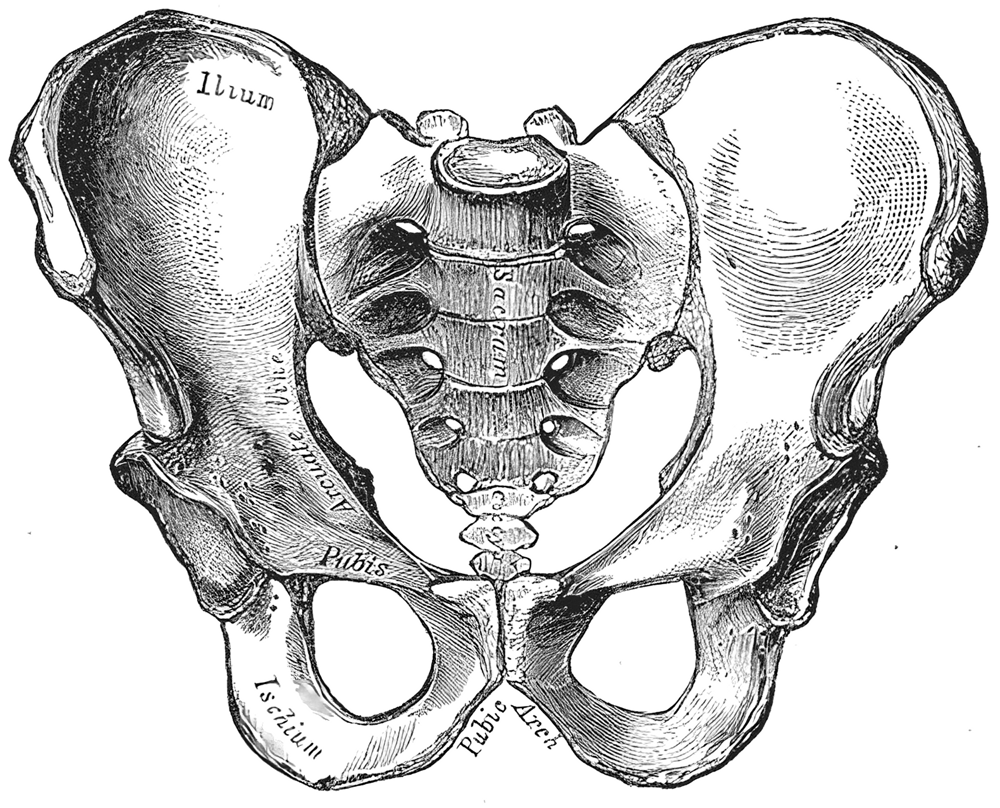


Anatomical components
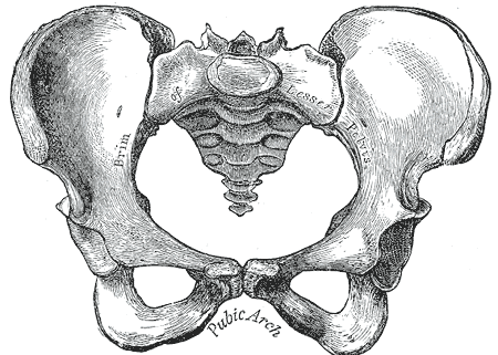
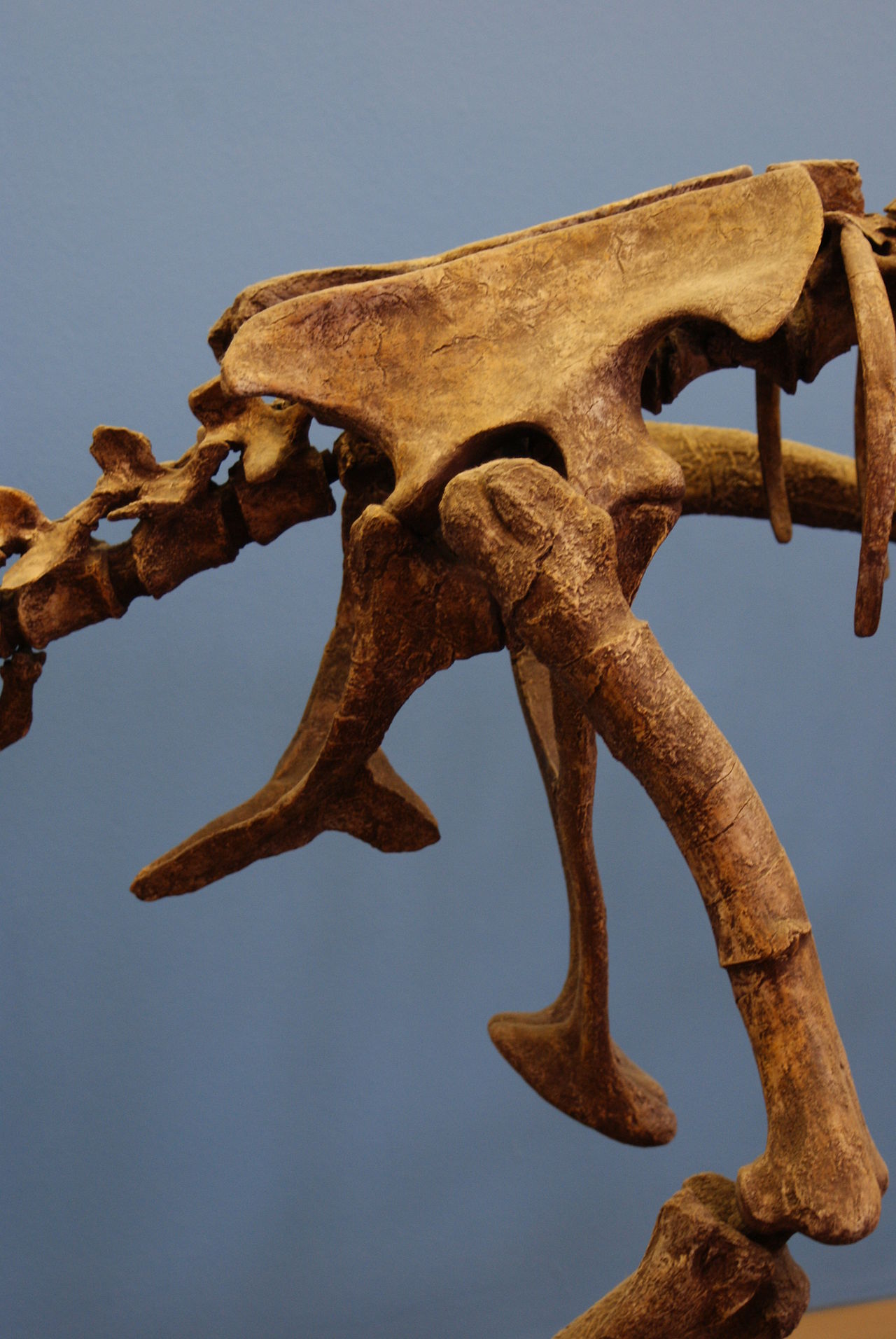
The adult pelvic skeleton is composed of three main elements that are fused in maturity: the paired hip bones (each often called the os coxae), the sacrum and the coccyx. At the rear the sacrum and coccyx form the connection to the spine; see spine, sacrum and coccyx. Each hip bone itself develops from three regions:
- Ilium — the broad, flaring superior part (ilium).
- Pubis — the anterior portion that joins at the midline.
- Ischium — the posteroinferior portion that bears weight in sitting.
These bones join at the sacroiliac joints, at the pubic symphysis and by strong ligaments that stabilise the ring. The hip socket (acetabulum) is formed where these regions meet and receives the femoral head to create the hip joint.
Functions and relationships
The pelvis performs multiple roles: it transfers weight from the trunk to the lower limbs during standing and locomotion; it protects pelvic and lower abdominal organs including parts of the digestive and reproductive systems; and it provides numerous sites for muscle and tendon attachment which control posture and movement. For its protective role with abdominal organs see abdomen and for digestive relations see digestive structures.
Development, variation and evolution
During growth the three parts of each hip bone are separate and later ossify and fuse. Human pelvic form reflects evolutionary adaptations to upright posture, bipedal gait and, in females, to childbirth. Pelvic bones are generally symmetrical left-to-right, although individual variation exists; symmetry is discussed in anatomical texts (symmetry).
Sex differences and clinical notes
Typical differences between female and male pelvises concern the shape and dimensions of the pelvic inlet and outlet, the angle of the pubic arch and the relative breadth of the ilia; these reflect obstetric requirements in females. Clinically, the pelvis is relevant for fractures, degenerative joint disease, sacroiliac dysfunction and obstetric assessment. The hip bone as a unit is sometimes referred to simply as the hip bone, and its subdivisions and landmarks are important in surgery, radiology and forensic identification.
Further reading and resources
Because the pelvis intersects many specialties — orthopaedics, obstetrics, anatomy and evolutionary biology — introductory and specialised references are useful for deeper study. General anatomical atlases and clinical guides cover development, biomechanics and variations in greater detail; an introductory note on morphology is available at vertebrate body and regional summaries may be found via institutional resources (thighs, abdomen, spine, sacrum, coccyx, digestive, symmetry, hip bone, ilium).
Questions and answers
Q: What is the pelvis?
A: The pelvis is the part of the vertebrate body to which the legs attach. It is located at the lower end of the spine and protects organs used for digestion and reproduction.
Q: What are the three bones that make up a hip bone?
A: The three bones that make up a hip bone are the ilium, pubis, and ischium.
Q: What other two bones make up the pelvic skeleton?
A: The sacrum and coccyx also make up part of the pelvic skeleton.
Q: How are muscles attached to bones in this region?
A: Muscles in this region are attached to bones with tendons.
Q: How do ligaments connect these bones together?
A: Ligaments connect these bones together by attaching them to each other and to the sacrum.
Q: Are these bones symmetrical on both sides?
A: Yes, these bones are symmetrical on both sides.
Related articles
Author
AlegsaOnline.com Pelvis (human anatomy): structure, function and development Leandro Alegsa
URL: https://en.alegsaonline.com/art/75513
Sources
- ncbi.nlm.nih.gov : ncbi.nlm.nih.gov/pmc/articles/PMC1997320/