Optic tract (visual pathway segment)
The optic tract is a paired bundle of nerve fibers in the brain that relays information from the eyes to central visual nuclei; it carries signals representing the contralateral visual field and is clinically important.
Overview
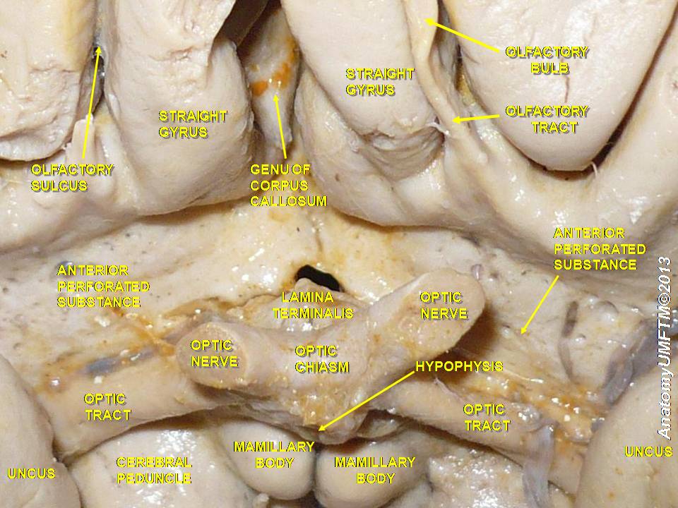
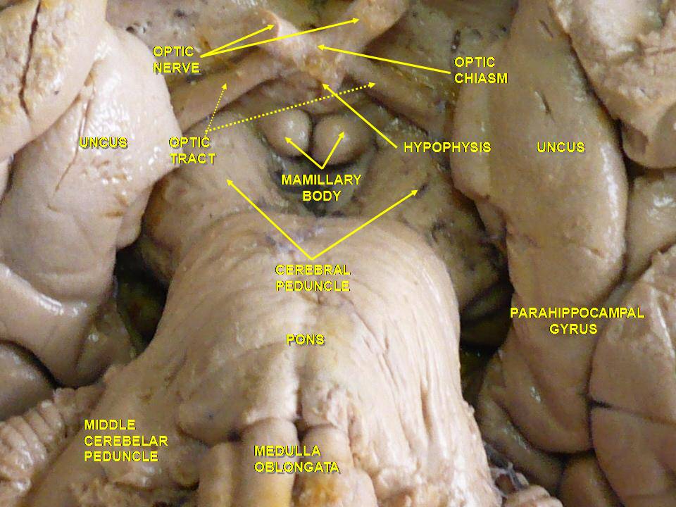
The optic tract is a major conduit in the visual system of the brain. It begins at the optic chiasma, where the two optic nerves meet and some fibers cross to the opposite side. From the chiasma, axons travel posteriorly as the left and right optic tracts to reach central visual processing centers. Together these tracts transmit condensed, organized information from the retina toward nuclei that analyze pattern, motion and reflexive responses.
Image gallery
10 Images


Organization and fibers
Each optic tract carries information about the contralateral half of the visual field. This arrangement arises because nasal retinal axons cross at the chiasm while temporal retinal axons remain on the same side. Thus the left optic tract conveys visual input from the right visual field and the right tract conveys the left field. The contributing axons are retinal ganglion cell fibers; in anatomical descriptions they are often referred to as retinal fibers.
Connections and functions
- Primary relay: most optic tract axons terminate in the lateral geniculate nucleus (LGN) of the thalamus, which forwards visual information to the visual cortex.
- Subcortical targets: branches also project to the superior colliculus and pretectal areas to support eye movements, spatial orientation and the pupillary light reflex.
- Integration: the tract groups input by visual field rather than by eye, facilitating binocular visual processing in downstream structures.
Clinical significance and pathology
Lesions of an optic tract typically produce a homonymous hemianopia, a loss of the same half of the visual field in both eyes. Depending on exact location and extent, patients may show partial field deficits, relative sparing of central vision, or disturbances in reflexive pupil responses. Because the optic tract is part of the central nervous system, it can be affected by inflammatory or demyelinating disorders (for example multiple sclerosis), tumors, vascular injury or traumatic damage. Clinical assessment commonly uses visual field testing and neuroimaging to localize tract lesions.
History, development and notable facts
Understanding of the optic tract developed through classical anatomy and later through physiological experiments and modern neuroimaging. Embryologically, retinal ganglion cell axons extend toward and across the chiasm to establish the adult pattern of crossing and uncrossed fibers. A practical distinction is that these are central nervous system axons (myelinated by oligodendrocytes) rather than peripheral nerves, which has implications for disease and repair research.
For more technical detail or clinical protocols consult specialized neuroanatomy and ophthalmology texts or review articles available through clinical resources: visual pathway overview, brain anatomy resources, optic chiasm references, optic nerve material and retinal connectivity summaries.
Questions and answers
Q: What is the optic tract?
A: The optic tract is a part of the visual system in the brain.
Q: Where does the optic tract start?
A: The optic tract starts from the optic chiasma.
Q: What does the optic chiasma do?
A: The optic chiasma receives the optic nerves from the eyes.
Q: How many optic tracts are there?
A: There are two individual tracts - the left optic tract and the right optic tract.
Q: What does each optic tract pass visual information from?
A: Each optic tract passes visual information only from the other side of the visual field.
Q: How does each optic tract develop?
A: Each optic tract develops from retinal fibers from each eye that corresponds to one half of the visual field.
Q: What is the function of the optic tract?
A: The optic tract is responsible for carrying visual information from the optic chiasma to the visual cortex in the brain.
Related articles
Author
AlegsaOnline.com Optic tract (visual pathway segment) Leandro Alegsa
URL: https://en.alegsaonline.com/art/72884
Sources
- britannica.com : britannica.com/science/optic-tract