
Nuclear magnetic resonance (NMR)
NMR is a physical phenomenon and analytical technique in which nuclei with nonzero spin absorb and re-emit radiofrequency energy in a magnetic field; it underpins spectroscopy, structural analysis and MRI.
Overview
Nuclear magnetic resonance (NMR) describes the interaction of atomic nuclei that possess a magnetic moment with an external magnetic field. In this field certain nuclei absorb and then re-emit electromagnetic radiation at characteristic frequencies. The absorbed energy corresponds to transitions between quantized nuclear spin states and depends on the applied magnetic field strength and on the magnetic properties of the isotopes involved. Small variations in the local electronic environment shift these resonance frequencies and form the basis of chemical analysis by NMR spectroscopy.
Image gallery
7 Images


Basic principles
NMR signals originate from nuclei with nonzero spin such as 1H, 13C, 15N and 31P. The resonance frequency for a given nucleus is proportional to the external field and the nucleus-specific gyromagnetic ratio. In molecules, the observable features include chemical shifts (sensitive to electronic shielding), spin–spin coupling (through-bond interactions) and relaxation times (T1 and T2) that report on molecular motion and local environment.
Instrumentation and methods
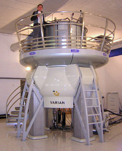
Modern NMR instruments use strong, stable superconducting magnets together with radiofrequency transmitters and sensitive receivers. Experiments may be continuous-wave or, more commonly today, pulsed with Fourier-transform detection. Pulse sequences are tailored to emphasize particular information—connectivity, distance constraints, diffusion or dynamics. Solid-state approaches adapt the method for powders and crystals, while solution-state techniques are optimized for molecules in liquid media.
Applications
NMR spectroscopy is a central tool in chemistry, biochemistry and materials science for determining molecular structures, conformations and interactions. It is non-destructive and quantitative, often requiring minimal sample preparation. The same physical principles are applied at different scales in magnetic resonance imaging (MRI), which produces detailed images of soft tissues for medical diagnosis. Additional applications include metabolomics, process monitoring and studies of molecular dynamics.
Limitations and developments
Key limitations of NMR include its relatively low intrinsic sensitivity compared with some other spectroscopic methods and the need for strong magnets and careful calibration. Only nuclei with nonzero spin are directly observable, which restricts the set of detectable elements. Ongoing developments—higher-field magnets, cryogenic probes, hyperpolarization methods and improved pulse sequences—seek to enhance sensitivity and broaden the range of practical experiments.
For introductory and advanced material, consult textbooks, review articles and technical notes from instrument manufacturers and academic groups. Practical resources and protocols are widely available through technical literature and specialist online collections.
History and development
Before 1940: Zeeman effect and Rabi method
In 1896 it was discovered that optical spectral lines split in the magnetic field (Zeeman effect). Hendrik Antoon Lorentz interpreted this soon after that the (circular) frequency of the light wave shifts by the amount of the Larmor frequency  because the atom represents a magnetic gyroscope which is excited by the magnetic field to a precession motion with the Larmor frequency.
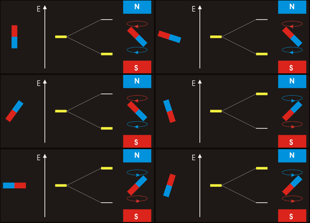
According to the light quantum hypothesis (Einstein 1905), the frequency shift by corresponds to an energy change Δ  , which in turn could be explained by the directional quantization of angular momentum discovered by Arnold Sommerfeld in 1916. With the angular momentum vector, the magnetic dipole of the atom, which is parallel to it, also has only discrete allowed setting angles to the magnetic field and correspondingly different discrete values of the magnetic energy. Thus the magnetic field causes the splitting of an energy level into several so-called Zeeman levels. This picture was directly confirmed in 1922 in the Stern-Gerlach experiment. There it was shown that the smallest possible (non-vanishing) angular momentum (i.e. quantum number
  ) can have only two possible setting angles to an external field.
At the end of the 1920s it was discovered that atomic nuclei have a magnetic moment about 1000 times smaller than atoms, which is why the splittings of energy levels caused by them are called hyperfine structure. The transition frequencies between adjacent hyperfine levels are in the range of radio waves (MHz). In 1936, Isidor Rabi succeeded in experimentally demonstrating that the precessional motion of atoms flying in an atomic beam through a constant magnetic field is disturbed by the irradiation of an alternating magnetic field when its frequency resonates with such a transition frequency. As a result, the magnetic moments of numerous nuclei could be determined with high accuracy, which, among other things, enabled the development of more accurate nuclear models.
1940s: Nuclear magnetic resonance in liquids and solids
Nuclear spin resonance in the narrower sense, i.e. the change in the angle of the nuclear spins with respect to the static external magnetic field without any significant participation of the atomic shell in the precession motion, was first realized in 1946 in two different ways. Edward Mills Purcell used the transfer of energy from the alternating magnetic field to the nuclear spins and further into their atomic environment to demonstrate resonance. Felix Bloch observed the alternating voltage induced by the preceding dipole moment of the nuclei in a coil when this is no longer parallel to the direction of the static field in the resonance case (method of "nuclear induction"). The prerequisite is that the static magnetic field causes the strongest possible polarization of the nuclear spins, which has oriented the development of devices towards ever stronger magnetic fields (today with superconducting coils up to 24 Tesla). These methods now enabled measurements on liquid and solid matter and a further increase in measurement accuracy to soon 6-8 decimal places. Accordingly, the measured values for the nuclear magnetic moments obtained with these methods were accurate. In a reversal of the problem, nuclear magnetic resonance thus also became a common method for the precision determination of magnetic fields. Moreover, various additional influences of the atomic environment on the magnetic field acting at the site of the nuclei became measurable, which, although small, allow detailed conclusions to be drawn about the structure and bonding ratios of the molecules and their mutual influence. Therefore, nuclear magnetic resonance spectroscopy is still a standard method in chemical structure research and one of the most important tools in analytical organic chemistry.
Applications in chemistry were initially considered unlikely. Among the pioneers was Rex Edward Richards in England, who was supported by Linus Pauling in not listening to skeptics. In Felix Bloch's group, Martin Everett Packard first recorded the NMR spectrum of an organic molecule in 1946. A breakthrough for the commercial market of using NMR spectrometers in organic chemistry was the NMR spectrometer A-60 from Varian Associates, developed in 1961 by James Shoolery at Varian, who also did as essential work in spreading knowledge of NMR among chemists and in popularizing it. Another pioneer of NMR spectroscopy in organic chemistry was John D. Roberts.
1950s: High-frequency pulses and spin echo
The measurement possibilities of the nuclear induction method expanded in the 1950s, when the use of the 10-20 MHz alternating field in the form of short pulses made it possible to manipulate the direction of the polarization of the nuclei. If the polarization is initially parallel to the constant magnetic field, for example, a "90° pulse" can be used to rotate the entire dipole moment of the sample in a specific direction perpendicular to the field direction. This allows direct observation of the subsequent free Larmor precession of the dipole moment about the field direction, because it induces (like the rotating magnet in an electrical generator) an alternating voltage in an antenna coil ("free induction decay", FID). The amplitude then decreases in time because the degree of alignment of the nuclear spins along the common direction perpendicular to the field decreases, partly because the polarization parallel to the static magnetic field is restored (longitudinal relaxation), partly because of field inhomogeneities and fluctuating interference fields (transverse relaxation). Both processes can be observed separately here, especially by means of the spin-echo method first described by Erwin Hahn.
1970/80s: NMR tomography and imaging
From the 1970s onwards, based on the work of Peter Mansfield and Paul C. Lauterbur, nuclear magnetic resonance was further developed into an imaging method, magnetic resonance imaging. When a strongly inhomogeneous static field is applied, the resonance frequency becomes dependent on the location of the nuclei in a controlled manner (field gradient NMR), but only in one dimension. From this, a three-dimensional picture of the spatial distribution of the nuclei of the same isotope can be obtained if the measurements are repeated successively with different directions of the inhomogeneous static fields. To obtain an image as rich in information as possible, e.g. for medical diagnoses, not only the measured values for the concentration of the isotope in question are then used, but also those for the relaxation times. These devices use superconducting magnets and 400 to 800 MHz alternating fields.
Special developments
Of principal physical interest are two more rarely used methods:
- As early as 1954, it was possible to detect the Larmor precession of the hydrogen nuclei (protons) of a water sample in the Earth's magnetic field (approx. 50 μT) using the FID method. The protons had been polarized by a stronger field perpendicular to the Earth's field, which had been rapidly switched off at a certain time. The immediate onset of Larmor precession induces an alternating voltage with a frequency of about 2 kHz, which is used, for example, to accurately measure the Earth's magnetic field. Absorption from a resonant alternating field is not required here. Therefore, this is the purest case of observing nuclear induction.
- Nuclear spin resonance has been successfully demonstrated on nuclei in a sufficiently long-lived excited state (shortest lifetime so far 37 μs), using for the detection here the modified angular distribution of the γ-rays emitted by the nuclei.
Physical basics
In the case of nuclear spin resonance, macroscopic explanations according to classical physics and microscopic explanations according to quantum mechanics can be easily combined (more detailed justification here). The decisive factor is that the Larmor precession of the nuclear spins has a magnitude and direction independent of their orientation. The corresponding effect of the static field can thus be completely transformed away by transition to a reference frame rotating about the field direction with the Larmor frequency, independent of the respective state of the individual nuclei considered in the sample and the magnitude and direction of the macroscopic magnetic moment formed by them.
Polarization
A nucleus with magnetic moment μ  has potential energy
  depends on the angle
 . The lowest energy belongs to the parallel position of the momentum with respect to the field, and the highest energy applies to antiparallel setting. In thermal equilibrium at temperature ,
  the moments are distributed among the different energies
  according to the Boltzmann factor ( }/k_{\mathrm
  ) . For typical nuclear moments
  and typical thermal energies
 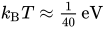 the Boltzmann factors differ by only less than 10-4, but the statistical preference for the small setting angles over the large ones is expressed by a nonzero mean ⟨.
  There arises a polarization and hence a macroscopic magnetic moment
 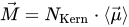 parallel to the external field (in which
  : number of nuclei). So much for the classical explanation of polarization by (nuclear) paramagnetism.
Zeeman levels
→ Main article: Zeeman effect
According to quantum mechanics, in states with definite angular momentum, any vector operator acts in parallel with the angular momentum operator  , one writes
 .
.
The constant γ  is called the gyromagnetic ratio, it has a characteristic value for each nuclide (see also Landé factor).
For the vector μ therefore also the directional quantization known from the angular momentum is valid, according to which, for a given angular momentum quantum number  , the cosine of the setting angle to the field direction in the energy eigenstates can only assume the values
  where the magnetic quantum number has the values
  . The largest possible component of μ along the field, also called the magnitude of the magnetic moment, is therefore μ
  .
.
Consequently, the component μ  of the moment parallel to the field has one of the values
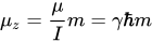
and the magnetic energy  accordingly:

(  : magnitude of .) This formula gives the energies of the
  Zeeman levels resulting from the equidistant splitting of the level with nuclear spin . The distance of adjacent Zeeman levels is just equal to the Larmor frequency ω
  , i.e., the frequency at which a (classical as well as quantum) magnetic gyroscope
  precesses in the field
 .
.
The occupation numbers of Zeeman levels decrease in thermal equilibrium from  to
to  (for positive γ , otherwise vice versa), but by no more than 10-4 relative in magnitude.
Relaxation
→ Main article: Relaxation (NMR)
The adjustment of the equilibrium polarization of the nuclear spins parallel to the external field is called longitudinal relaxation. It takes up to several seconds in liquid and solid samples (in gases it can take weeks), if the sample does not contain paramagnetic admixtures, i.e. atoms with permanent magnetic dipole moment, which cause transitions between the Zeeman levels by fluctuating magnetic fields and thus accelerate the energy exchange with the nuclear spins. The time constant is denoted by  . The decay of a polarization perpendicular to the field to zero equilibrium value is called transverse relaxation and is (usually) faster (time constant
  ), because no energy turnover is required for this; rather, it is sufficient that the nuclear spins aligned transverse to the magnetic field lose their common alignment due to small fluctuations in their continuous Larmor precession about the field direction. Temporally, the approach to equilibrium follows a good approximation of a simple decaying exponential function.
Bloch equations
→ Main article: Bloch equations
The Bloch equations summarize Larmor precession and longitudinal and transverse relaxation in a single equation of motion for the vector of magnetic moment  together (with magnetic field
 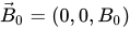 and equilibrium magnetization
 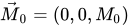 , both parallel to the
  -axis):
![{d \vec M \over dt} = \gamma \vec M \mathord \times \vec B_0\ - \ \begin{pmatrix} \tfrac{M_x }{T^*_2} \\[0.5em] \tfrac{M_y }{T^*_2}\\[0.5em] \tfrac{M_z-M_0 }{T_1} \end{pmatrix}](https://www.alegsaonline.com/image/a30975220685c81de5ef3c633b60e2889b427c85.svg)
In it, the cross product describes the Larmor precession with angular velocity ω  . In the 2nd term, the relaxation is phenomenologically summarized as a 1st order process (i.e., simple exponential decay), where the time constant for the component of
  parallel to the field is different from that for the transverse ones. According to quantum mechanics, the Bloch equations also hold for the expectation value of the magnetic moment of each nucleus ⟨
Transverse alternating field and absorption of energy
A weak additional alternating field, e.g., in the  direction, can always be conceived as the sum of two circularly polarized alternating fields rotating, e.g., about the axis (i.e., the direction of the strong constant field) in opposite senses.
- In quantum mechanical terms, this alternating field induces transitions between Zeeman levels in one direction or the other in the resonance case, because its circularly polarized quanta have the right angular momentum ( component
 ) and with
  then just the right energy. These transitions disturb the thermal equilibrium because they reduce existing differences in occupation numbers. This implies a net energy absorption because more nuclei were previously in lower energy states than in higher ones, according to the thermal equilibrium. This flow of energy from the alternating field into the system of nuclear spins would come to a halt when equal occupation was reached. The thermal contact of the spin system to the environment, which is already decisive for the production of the original equilibrium magnetization, continuously withdraws energy from the thus disturbed spin system. A steady-state equilibrium is reached with a slightly reduced magnetization. The relevant parameter is the longitudinal relaxation time . The first proofs and applications of nuclear magnetic resonance according to Purcell's method are based on this continuous wave method.
 - In macroscopic view it is easier to overlook which movement of the macroscopic dipole moment results from it: In the resonance case, the two components of the alternating field co-rotating with the Larmor precession represent a constant field perpendicular to the -axis. It acts on the dipole with a torque which imposes on it a further Larmor precession about the axis (co-rotating in the xy-plane) of this additional field. Since in this process the setting angle to the much stronger static field must change, the dipole absorbs or releases energy from the alternating field. If the dipole was previously parallel to the field direction , it can induce an alternating voltage in a receiver coil when twisted itself. If the alternating field is pulsed, depending on the exposure time, the dipole moment can, for example, be specifically rotated exactly 90° or even completely reversed (as far as the relaxation time allows). This gives rise to the numerous different pulse methods with their versatile measurement possibilities (e.g., the spin echo for the separate determination of and ).
Related articles
Author
AlegsaOnline.com Nuclear magnetic resonance (NMR) Leandro Alegsa
URL: https://en.alegsaonline.com/art/71361
Sources
- ui.adsabs.harvard.edu : 1938PhRv...53..318R
- doi.org : 10.1103/PhysRev.53.318
- pubmed.ncbi.nlm.nih.gov : 9981980
- nobelprize.org : Biography of I. Rabi at Nobelprize.org
- doi.org : 10.1038/npre.2009.3267.5
- nobelprize.org : 1952 Nobel Prize for Physics at Nobelprize.org