Atomic force microscope
An overview of the atomic force microscope (AFM): principles, main components, imaging modes, history, common applications and limitations in nanoscience and biology.
Overview

An atomic force microscope (AFM) is a high‑resolution scanning probe instrument used to measure and image surfaces at the nanometre and, in favorable cases, atomic scale. Rather than relying on lenses or an electron beam, an AFM senses the topography and local properties of a sample by moving a very sharp tip over the surface and recording the interaction forces. AFMs are widely used in materials science, surface physics, chemistry, and biology and are an important tool in nanotechnology; for a general comparison with other instruments see microscopes and the specific contrasts with the scanning electron microscope. AFMs can operate in air, vacuum or liquid environments and so are particularly useful for imaging delicate or hydrated samples often encountered in biological research and soft materials.
Image gallery
10 Images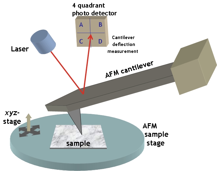


Main parts and operating principle
An AFM obtains surface information through a sharp probe mounted on a flexible cantilever. Key components include:
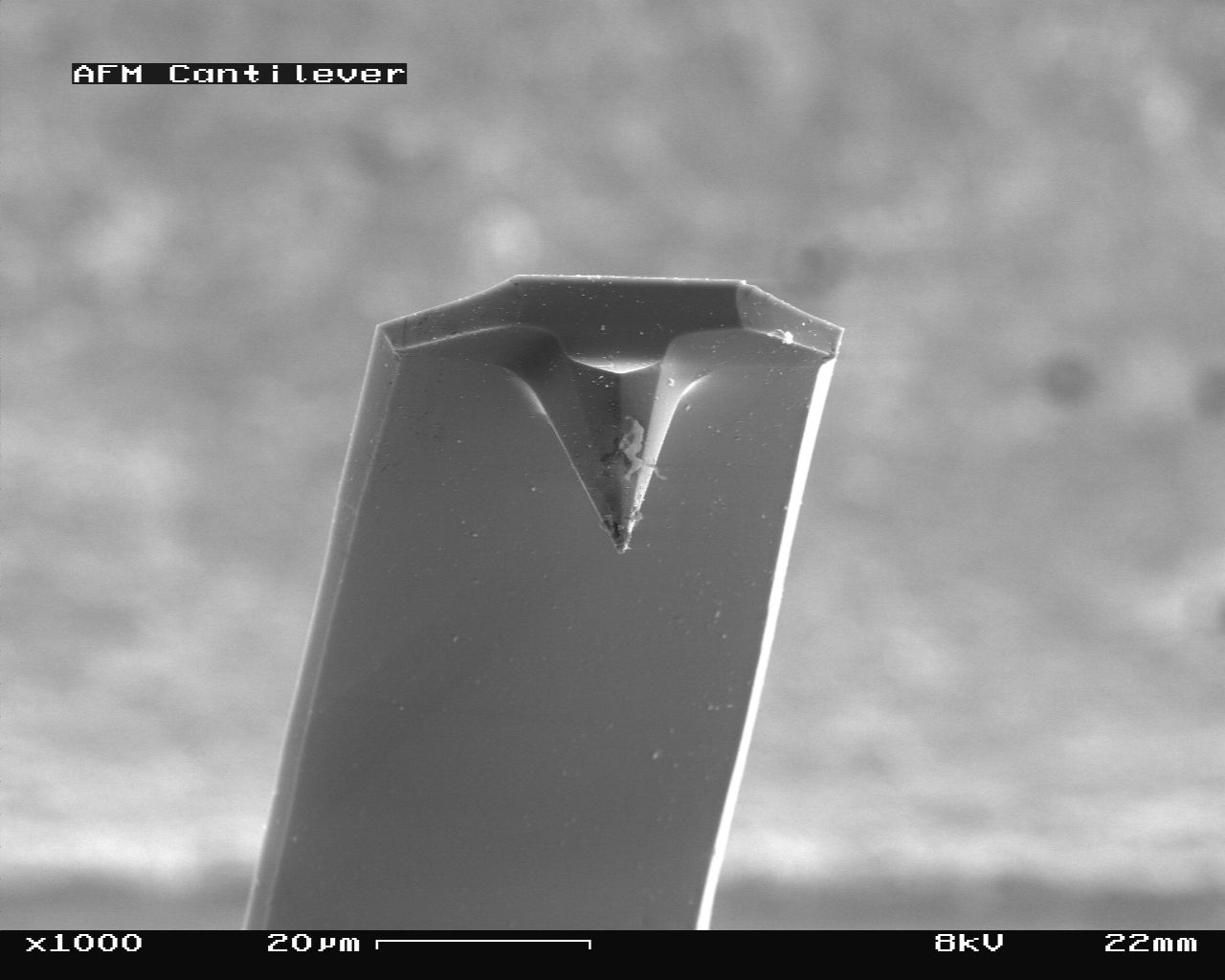
- Tip and probe: a nanometre‑scale sharp tip (often made of silicon or silicon nitride) at the end of a cantilever that interacts with the sample surface.
- Cantilever: a small beam that bends in response to forces between tip and surface; its deflection is used to infer surface features and forces.
- Deflection detector: commonly a laser beam reflected from the cantilever onto a segmented photodiode, which converts bending into an electrical signal.
- Scanner: a piezoelectric stage that raster‑scans the sample beneath the tip with subnanometre precision.
- Control electronics and software: closed‑loop systems maintain feedback and convert detector signals into height maps, force curves and other data.
Imaging modes and techniques
AFMs can be operated in several modes that emphasize different tip–sample interactions:
- Contact mode: the tip remains in continuous contact with the surface while lateral deflection maps topography. It offers straightforward height imaging but can damage soft samples.
- Tapping or intermittent contact mode: the cantilever oscillates and the tip periodically taps the surface. Feedback maintains a set oscillation amplitude; this reduces lateral forces and is gentler on biological or soft polymer samples.
- Non‑contact mode: the cantilever oscillates above the surface and senses long‑range attractive forces. This mode reduces tip wear and sample disturbance but can be more challenging in ambient conditions.
- Force spectroscopy and advanced modes: by measuring force versus separation, AFMs quantify mechanical properties such as stiffness, adhesion and viscoelasticity. Many instruments combine imaging with electrical, magnetic or thermal sensing.
History and development
The AFM grew out of the scanning probe microscope family developed in the 1980s. It was introduced to extend scanning tunnelling microscopy methods to insulating surfaces by sensing mechanical forces rather than tunnelling current. Since its invention, instrument design and tip fabrication have advanced substantially, enabling higher resolution, improved stability and operation in diverse environments. Research over subsequent decades produced many variants and specialized modes used for nanoscale manipulation, lithography and sensing.
Applications and significance
Atomic force microscopes are versatile research tools. Typical uses include imaging surface topography at nanometre resolution, measuring mechanical properties of thin films and biological specimens, characterizing nanoscale electronic or magnetic domains, and manipulating nanoparticles or molecules. AFM is routinely used in semiconductor R&D, polymer science, biomaterials and cell mechanics. Its ability to work in liquids makes it invaluable for studying membranes, proteins and living cells in near‑physiological conditions. For connections to applied fields see resources on nanotechnology.
Limitations and notable considerations
Despite its strengths, AFM has limitations. Scan speeds are typically slower than optical methods, images can be affected by tip shape (tip convolution), and delicate samples may be altered by tip forces if modes are not chosen carefully. Accurate quantitative measurements require calibration and careful control of environmental factors such as vibration, temperature and contamination. Nonetheless, when used appropriately, AFM provides unique, high‑resolution insights into surface structure and nanoscale properties that complement other microscopy techniques.
Questions and answers
Q: What is an atomic force microscope (AFM)?
A: An atomic force microscope (AFM) is a type of microscope that provides pictures of atoms on or in surfaces. It can be used to look at individual atoms and is commonly used in nanotechnology.
Q: How does the AFM work?
A: The AFM works by employing an ultra-fine needle attached to a cantilever beam. The tip of the needle runs over the ridges and valleys in the material being imaged, "feeling" the surface. As the tip moves up and down due to the surface, the cantilever deflects. In one basic configuration, a laser shines on the cantilever at an oblique angle, allowing for direct measurement of deflection in the cantilever by changing its angle of incidence for the laser beam. This creates an image revealing configuration of molecules being imaged by machine.
Q: What are some advantages that AFMs have over scanning electron microscopes (SEMs)?
A: AFMs provide higher resolution than SEMs and do not need to operate in a vacuum like SEMs do - they can operate in ambient air or water, allowing them to be used with biological samples such as living cells without damaging them.
Q: What are some operating modes for AFMs?
A: Commonly used operating modes for AFMs include contact mode, where tip is simply moved across surface and cantilever deflections are measured; tapping mode, where tip is tapped against surface as it travels along; intermittent contact mode; non-contact mode; dynamic mode; static mode; and more - these are often variations on tapping and contact modes described above.
Q: How does tapping mode differ from contact mode?
A: Tapping mode differs from contact mode because when using tapping mode, tip taps against surface as it travels along instead of just moving across it - this allows it to move away from surface when needle feels ridge so that it will not hit against surface when moving across which makes it useful for soft surfaces such as biological samples since less likely to damage them this way.
Related articles
Author
AlegsaOnline.com Atomic force microscope Leandro Alegsa
URL: https://en.alegsaonline.com/art/7047