Nervous tissue
The nervous tissue is one of the four basic tissues of tissue animals, which include, among others, vertebrates and so also humans. It consists of nerve cells and glial cells, both of which originate from common neuroectodermal precursor cells, which in chordates arise from the neural tube and the neural crest.
Nerve cells, or neurons, and glial cells together form the nervous tissue and interact to develop the basic structures of the nervous system. The neural tube thus becomes the central area with brain and spinal cord, in the peripheral area nerves, nerve plexuses and ganglia are formed, together with enteric parts in the gastrointestinal tract. Neurons and glia work together to carry out the basic functions of this system, the transmission of neuronal excitations.
Neurons form projections for this purpose, which receive excitation from other cells (dendrites) or communicate their own excitation to other cells (neurite). At the points of excitation transmission (synapses), neurons are linked in this way to form interconnected chains, loops or circuits. Of particular importance for the functional connection of such neuronal networks of a nervous system are the contacts of neurons through which it is related to the rest of the body and its environment. These include, on the one hand, afferent contacts of certain neurons to sensory cells - which can be specifically changed by their environment - such as sensory cells (sensors) and, on the other hand, efferent contacts of certain neurons to motor cells - which can specifically change their environment - such as muscle cells or glandular cells (effectors). Neurons that mediate between sensory or motor parts in a promoting or inhibiting manner are called interneurons.
In early development, glial cells form basic structures to which young neurons can orient themselves by migrating or forming processes; subsequently, they stabilize processes and connections by means of a sheath and later allow particularly rapid conduction of excitation (saltatory) by means of multiple sheaths. In the more mature nervous system, they ensure, among other things, low-disturbance signal transmission and signal transduction, absorb released messenger substances, provide nutrients and are involved in the blood-brain barrier and, as ependymal cells, in the blood-liquor barrier, with which the nervous tissue is separated in a special way from the intravascular space of the supplying blood capillaries.
In living organisms, nerve tissue appears pink or light grey to whitish, with fine structural differences. In the so-called gray matter, the bodies of nerve cells predominate, whose accumulations in the central nervous system (CNS) are also referred to as nuclei, and in the peripheral mostly as ganglia. The white matter consists mainly of the projections of nerve cells, which appear bright as axons surrounded by glial cells in the case of myelin-containing nerve fibers and are often combined into conduction pathways, for example as central projection pathways, in the peripheral nervous system (PNS) usually called nerves.

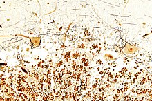
Tissue section from the cerebellum (Bielschowsky stain): In the image above, surrounded by glia stained light brown, the processes and large cell bodies of a few Purkinje cells are in close contact with some black-stained processes of basket cells. Below the position of the cerebellar granule cells; in mammals they represent more than 50 % of all neurons.
Regeneration of nerve tissue
The regenerative capacity of nerve tissue is very limited compared to other tissues, especially since nerve cells are no longer able to divide.
In early embryonic development, the nervous system anlage is for some time the region with the highest rate of cell divisions, and fetally, at peak times in humans, several thousand young neurons are produced per second. But these neurons are no longer capable of cell divisions afterwards, postmitotic. And not all of them live as long as the organ of the organism in whose tissue they seek their place (see selective apoptosis).
In the adult brain, only in a few regions undifferentiated neural precursor cells remain, which can continue to divide and are capable of forming neuroblasts and young neurons (see adult neurogenesis). In humans, for example, young neurons can also be formed alongside glial cells, for example in regions of the hippocampus or in the subventricular zone, to replace neurons in the olfactory bulb and olfactory mucosa. To do this, these young neurons must migrate into that brain region and search for a place (with chemotaxis or haptotaxis), extend extensions (axogenesis), form transmission sites (synaptogenesis), establish contacts in the found network of other neurons, receive signals and send signals, and finally also those with which the excitation state of certain individual other cells can be changed (excitation or inhibition).
On the way there and in the process along the way, a neuron differentiates - to take a place in a cellular environment with specific connections. If it does not succeed, the neuron does not survive for long. If it succeeds, the neuron occupies a special place in the neuronal network - and can only be replaced in this place by young neurons that follow a similar differentiation process. But these cannot be formed from mature neurons by cell division. For this, the neurons would have to round off, the processes would regress, lose their contacts and thus become non-functional. The replacement of differentiated and functional neurons within a neuronal network is therefore limited by the complexity of the neuronal connections.
In the peripheral nervous system, on the other hand, after damage to a nerve fiber, the extension of a neuron can grow back into the canal of the medullary sheath as an axon - if it is still present - at about the same rate as hair grows.
Search within the encyclopedia