
Nephron: Structure, Function, and Clinical Significance of the Kidney's Functional Unit
A clear overview of the nephron: its anatomy, physiological roles (filtration, reabsorption, secretion, concentration), types, development, and importance in health and disease.
Overview
The nephron is the microscopic functional unit of the kidney that performs blood filtration and urine formation. Each human kidney contains many hundreds of thousands to over a million nephrons; together they maintain fluid, electrolyte and acid–base balance and remove metabolic waste. Nephrons are embedded within the kidney's outer cortex and inner medulla and connect ultimately to the collecting system that drains into the renal pelvis and ureter (ureter), which conveys urine to the bladder. For general information about the organ that houses nephrons see the kidney overview at Kidney.
Image gallery
9 Images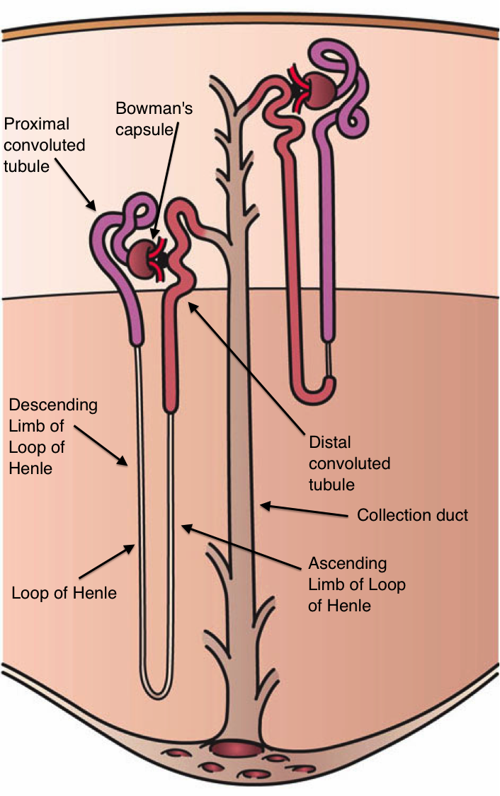


Anatomy and main parts
Each nephron is a continuous tubular structure associated closely with capillary networks. Major segments are:
- Renal (Bowman’s) capsule and glomerulus – a tuft of capillaries where plasma is filtered into the capsule space.
- Proximal tubule – bulk reabsorption of water, electrolytes and nutrients back into the blood.
- Loop of Henle – a hairpin-shaped segment that creates a concentrated medullary gradient important for water reabsorption.
- Distal tubule – fine-tuning of electrolyte and acid–base handling.
- Collecting duct – final adjustments in water and solute before urine leaves the nephron and flows into the collecting system.
Function and transport processes
Nephrons perform four core tasks: filtration, reabsorption, secretion and concentration. Blood pressure forces plasma through the glomerular filter into Bowman’s space; useful substances such as glucose and much of the filtered water are recovered in the proximal tubule; the loop of Henle and vasa recta set up a countercurrent multiplier and exchange system that enables the kidney to concentrate or dilute urine; the distal nephron and collecting duct perform regulated reabsorption and secretion under hormonal control (for example by aldosterone and antidiuretic hormone). The juxtaglomerular apparatus near the glomerulus senses blood flow and contributes to renal autoregulation and renin release, linking nephron function to systemic blood pressure control.
Types of nephrons and development
Nephrons fall into two general categories: cortical nephrons, with glomeruli in the outer cortex and short loops of Henle, and juxtamedullary nephrons, with glomeruli close to the medulla and long loops that are essential for concentrating urine. In human development nephrons arise from the intermediate mesoderm during formation of the permanent kidney (the metanephros). The total nephron number is largely determined before or shortly after birth; there is very limited capacity to generate new nephrons in adult humans, which has implications for recovery after injury or loss.
Clinical importance and examples
Because nephrons carry out filtration and fine control of body fluids, their loss or dysfunction causes predictable problems: reduced glomerular filtration rate (GFR) and impaired waste excretion, fluid overload or dehydration, electrolyte imbalances, and disturbances of acid–base homeostasis. Progressive nephron loss underlies chronic kidney disease; acute insults can cause sudden drops in function (acute kidney injury). Common diagnostic clues to nephron injury include protein or blood in the urine and changes in serum creatinine or electrolytes. Many pharmacologic agents act on specific nephron segments—for instance, loop diuretics target the thick ascending limb of the loop of Henle, and thiazide diuretics act on distal tubular transporters—to produce diuresis and alter electrolyte handling.
Notable facts and distinctions
Historically, the capsule around the glomerulus is known as Bowman’s capsule and the combined glomerular structure is sometimes called the Malpighian body after early anatomists. The number of nephrons varies widely among individuals and species; fewer nephrons can predispose to hypertension and renal disease. Because each nephron contributes only a small fraction of total renal function, substantial nephron loss can be clinically silent until compensation is exhausted.
In summary, the nephron is a compact but highly organized unit that couples vascular filtration to tubular modification of the filtered fluid. Its structure—specialized segments and close vascular relationships—allows precise control of body fluid composition and volume, making nephrons essential to homeostasis and human health.

Physiology
In the renal corpuscles, primary urine is continuously filtered from the blood. Subsequently, certain substances are reabsorbed in the tubules (especially water is "reabsorbed", reabsorbed, water reabsorption), but also secreted. Through this concentration, the primary urine becomes the actual urine (secondary urine or final urine). Primary urine formation is also called glomerular filtration, filtrative renal function, or creatinine clearance; it is about 150 liters per day (or 105 ml/min) in adults.
The tubules regulate the water balance, the glomeruli filter the plasma, each in proportion to the variable cardiac output.
The actual renal performance consists in the active transport of the tubules (under energy consumption) in contrast to the hemodynamically generated (passive) filtration of the glomeruli. The active transport processes in the renal tubules are divided into primary active, secondary active and tertiary active.
History
The theories of urine preparation have a long history. Leonhart Fuchs (1501-1566) already described the kidney as a sieve or filter. The Austrian anatomist Josef Hyrtl also referred to a kidney as a seihe (seyhe) or sieve. William Bowman claimed as late as 1842 that the glomerular capillaries secrete water, which washes away the substances secreted by the tubules. Carl Ludwig also first had a clear idea of how the kidneys work in the preparation of urine in 1842. According to his mechanical theory, which is still essentially valid today, the physical filtration of the plasma takes place in the glomeruli. Subsequently, water is re-diffused through endosmosis in the tubule.
The tubular reabsorption of urinary substances was not recognized until 1917 by Arthur Robertson Cushny. Today we speak of (passive, i.e. without energy consumption) glomerular filtration and (active, i.e. with energy consumption) tubular reabsorption. Already Franz Volhard rejected this "modern mechanical-physical filtration theory", although he described it correctly in detail several times ("Filtration-reabsorption theory of Ludwig and Cushny"). In 1934, Leopold Lichtwitz also considered even the possibility of filtration and ultrafiltration in the glomeruli to be impossible, because he could not imagine the passive passage of urinary substances through the slit membranes in the podocytes almost without energy consumption.
However, the interplay of physics and chemistry (neurohumorally regulated and drug-modulated) in health and disease in the podocytes and in the individual tubule sections with respect to the individual urinary and urinary substances has not been conclusively clarified even today.
Related articles
Author
AlegsaOnline.com Nephron: Structure, Function, and Clinical Significance of the Kidney's Functional Unit Leandro Alegsa
URL: https://en.alegsaonline.com/art/69188