
Myocyte (muscle cell): types, structure, development and function
Myocytes are the cells that make up muscle tissue. This article describes their types (skeletal, cardiac, smooth), structure, development (myogenesis), functions, regeneration and clinical relevance.
Overview
A myocyte, commonly called a muscle cell, is the basic cellular unit of muscle tissue. Myocytes are specialized for contraction and force generation through interactions between actin and myosin proteins. They arise during embryonic development from precursor cells and differentiate into several structurally and functionally distinct types that support movement, posture, circulation and internal organ function.
Image gallery
2 Images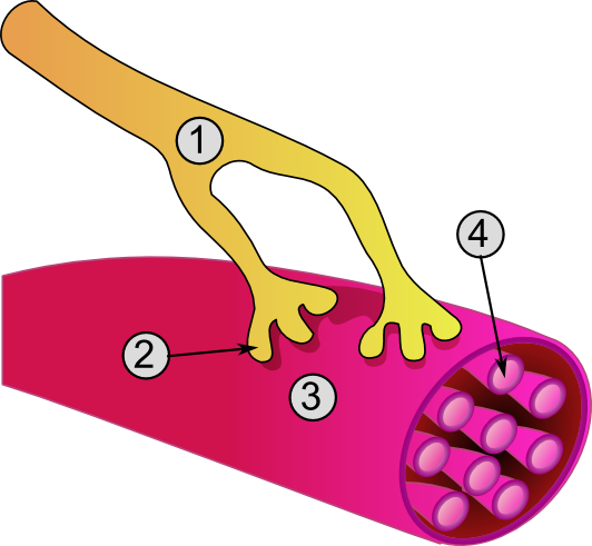

Major types and distinguishing features
There are three principal classes of myocytes, each adapted to particular roles:
- Skeletal myocytes are long, cylindrical and typically multinucleated. They show a repeating pattern of sarcomeres that produces visible striations and are under voluntary control for posture and locomotion. See skeletal muscle for more context.
- Cardiac myocytes form the working muscle of the heart. These cells are striated but usually have a single central nucleus, branch to form networks, and connect by specialised junctions that allow coordinated contraction. Related information is available at cardiac.
- Smooth muscle cells are spindle-shaped, non-striated, and typically have one central nucleus. They contract involuntarily to move contents through hollow organs and to regulate vessel diameter; see smooth muscle.
Structure and contraction mechanics
Myocytes contain contractile proteins arranged into organized units in striated cells (sarcomeres) or in a less-ordered network in smooth muscle. Contraction is driven by calcium-regulated interactions between actin and myosin filaments. Excitation–contraction coupling differs among types: skeletal muscle uses a neuromuscular junction and T-tubules, cardiac muscle relies on electrically coupled cells and calcium-induced calcium release, and smooth muscle uses a variety of signalling pathways responsive to nerves, hormones and mechanical stretch.
Development, growth and regeneration
Muscle cells originate from precursor myoblasts during a process called myogenesis, in which individual progenitor cells fuse or differentiate to form mature myocytes. Skeletal muscle retains a population of satellite cells that can divide and repair fibres after injury. Cardiac muscle has a limited capacity to regenerate in adults, and smooth muscle is comparatively better at proliferation and repair.
Functions and examples
Myocytes power diverse physiological activities: skeletal myocytes enable voluntary movement and breathing, cardiac myocytes pump blood throughout the body, and smooth muscle cells produce involuntary movements such as peristalsis in the digestive tract. Examples of smooth muscle action include coordinated contractions in the oesophagus and rhythmic churning in the stomach.
Clinical relevance and notable facts
Muscle cells are central to many diseases and clinical tests. Damage to cardiac myocytes releases biomarkers used to diagnose heart injury. Genetic disorders and inflammatory conditions can affect skeletal muscle strength and repair. Understanding differences among myocyte types is important in regenerative medicine, pharmacology and surgery. For concise summaries of their roles and distinctions, consult general references on skeletal, cardiac and smooth muscle.
Terminology note: Individual multinucleated skeletal cells are often called muscle fibres or myofibres; cardiac muscle cells are commonly termed cardiomyocytes. Though varying in shape and control mechanisms, all myocytes share the primary function of producing force through contraction.
Questions and answers
Q: What is a myocyte?
A: A myocyte is a type of cell found in muscle tissue.
Q: What is the process of myogenesis?
A: Myogenesis is the process by which myoblasts develop into myocytes to form muscles.
Q: What are the specialized forms of myocytes?
A: The specialized forms of myocytes are cardiac, skeletal, and smooth muscle cells.
Q: What is the structure of cardiac and skeletal muscle cells?
A: The striated or striped cells of cardiac and skeletal muscles are called muscle fibers.
Q: What are cardiomyocytes?
A: Cardiomyocytes are the muscle fibres which form the chambers of the heart.
Q: What is the difference between the nuclei of cardiomyocytes and skeletal muscle fibers?
A: Cardiomyocytes have a single central nucleus while skeletal muscle fibers usually have peripheral nuclei.
Q: What is the function of smooth muscle cells?
A: Smooth muscle cells control involuntary movements such as the peristalsis contractions in the oesophagus and stomach.
Related articles
Author
AlegsaOnline.com Myocyte (muscle cell): types, structure, development and function Leandro Alegsa
URL: https://en.alegsaonline.com/art/67982
Sources
- meshb.nlm.nih.gov : meshb.nlm.nih.gov/record/ui?name=Myocytes
- botany.uwc.ac.za : "Muscle tissues"
- pubmed.ncbi.nlm.nih.gov : 11694174
- ptjournal.apta.org : "Human skeletal muscle fiber type classifications" · web.archive.org
- researchgate.net : "Does anyone know why skeletal muscle fibers have peripheral nuclei, but the cardiomyocytes not? What are the functional advantages?"